Photoacoustic analyzer of region of interest in a human body

a photoacoustic analyzer and human body technology, applied in the field of medical devices and methods, can solve the problems of fetal death, non-reassurance of fhr always associated, inaccurate readings in the presence of ambient light, etc., and achieve the effect of improving the signal to noise ratio (snr) of the detection system and safer light energy for illumination

Inactive Publication Date: 2009-04-07
ORNIM
View PDF9 Cites 116 Cited by
  • Summary
  • Abstract
  • Description
  • Claims
  • Application Information

AI Technical Summary

Benefits of technology

[0015]Alternatively or additionally, the control unit is configured and operable to define a focal volume for collection of sound waves (acoustic radiation) to match dimensions of the region of interest, such that the acoustic unit collects a substantial portion of these sound waves during a predetermined time window Δt. It should be understood that such a matching region may be the entire region of interest or a portion thereof. This technique improves the signal to noise ratio (SNR) of the detection system, enabling use of safer light energies for illumination. The collected signals are processed and analyzed to determine the presence, and optionally, the amount or concentration, of a substance in the region of interest in the irradiated body tissues or fluids.

Problems solved by technology

However, it can provide inaccurate readings in the presence of ambient light, in cases of low perfusion of blood and during motion of the subject or the portion of tissue being examined.
A normal fetal-heart-rate (FHR) pattern is usually associated with the delivery of a normal well-oxygenated infant; however, a non-reassuring FHR is not always associated with the delivery of a compromised infant.
If the baby is delivered and the lungs are not mature, the baby may develop Respiratory Distress Syndrome (RDS), which can result either in fetal death or in long-lasting periods of repeated respiratory difficulty.
Although it is the clinical gold standard method, it remains a time-consuming process, has a large intralaboratory and interlaboratory coefficient of variation, and requires expertise.
In addition, the procedure of amniotic fluid drainage itself is invasive and suffers a small risk of abortion.
In cases where the fetus gasps during delivery, inhaling the sticky meconium into the upper respiratory tract results in partial airways obstruction.
However, in cases where the fetus head is tightly fitted in the pelvis, the amniotic fluid is not drained, preventing detection of any potentially harmful outcome to the respiratory tract.

Method used


Image

  • Photoacoustic analyzer of region of interest in a human body
  • Photoacoustic analyzer of region of interest in a human body
  • Photoacoustic analyzer of region of interest in a human body

Examples


Embodiment Construction

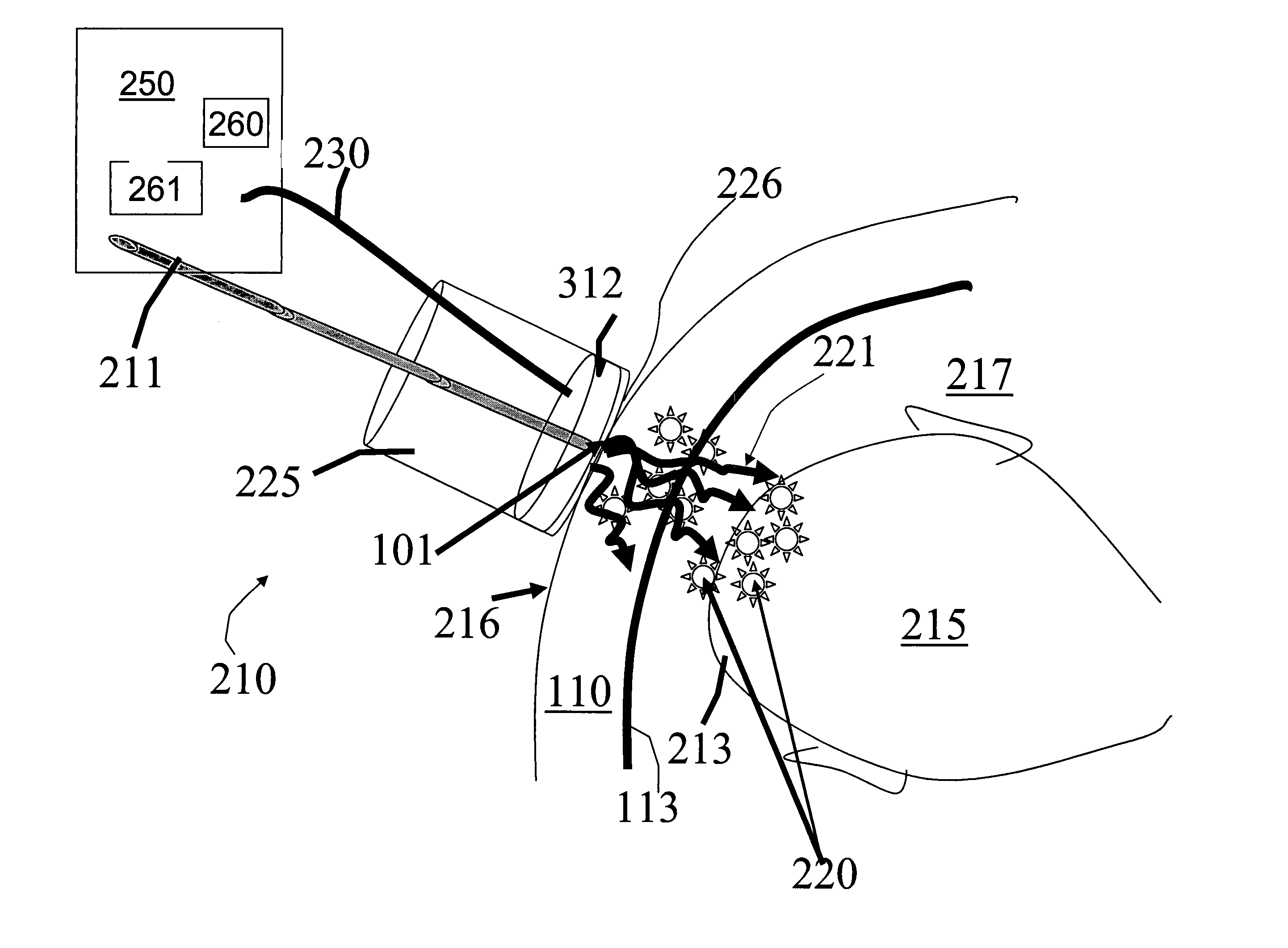
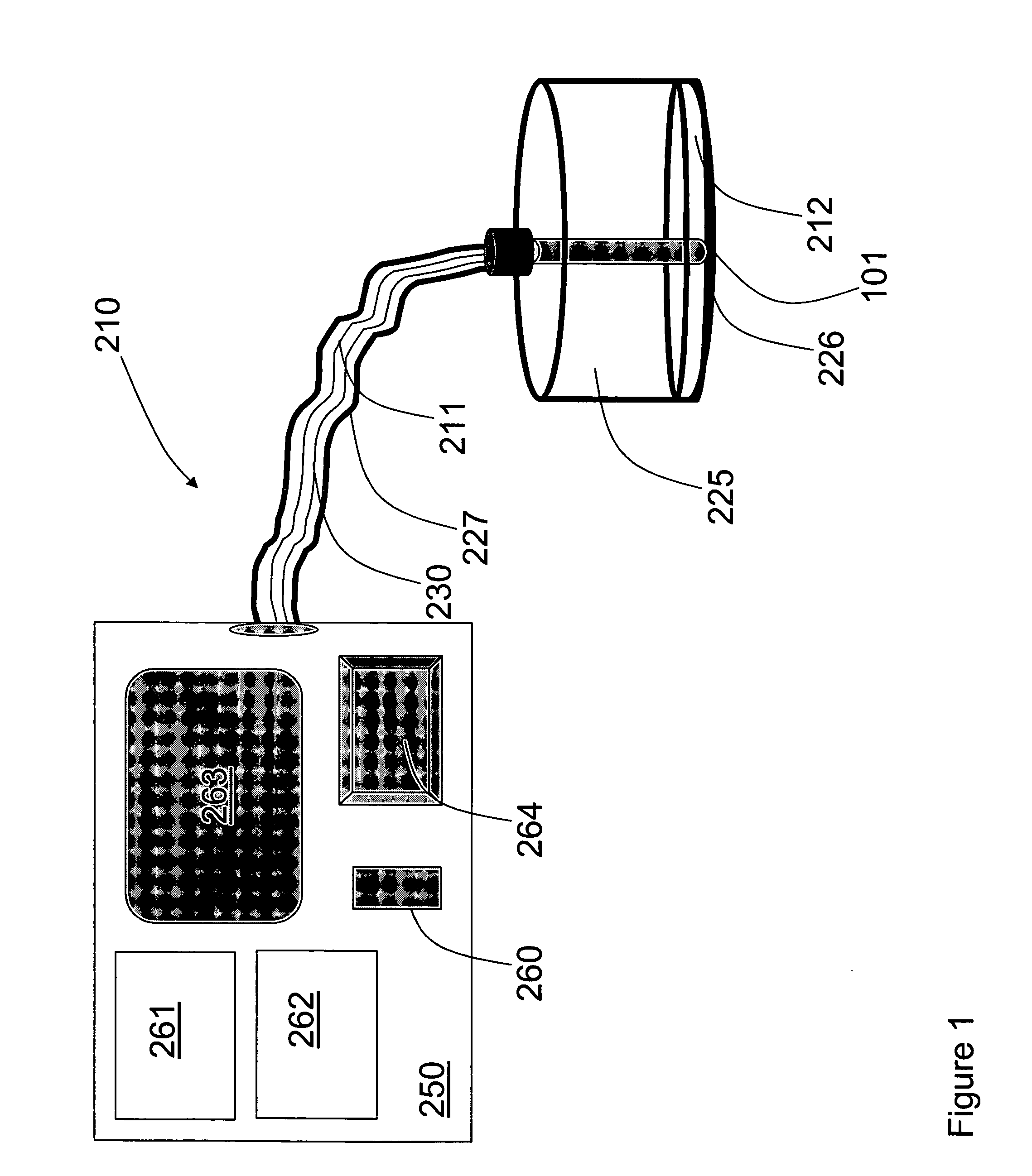
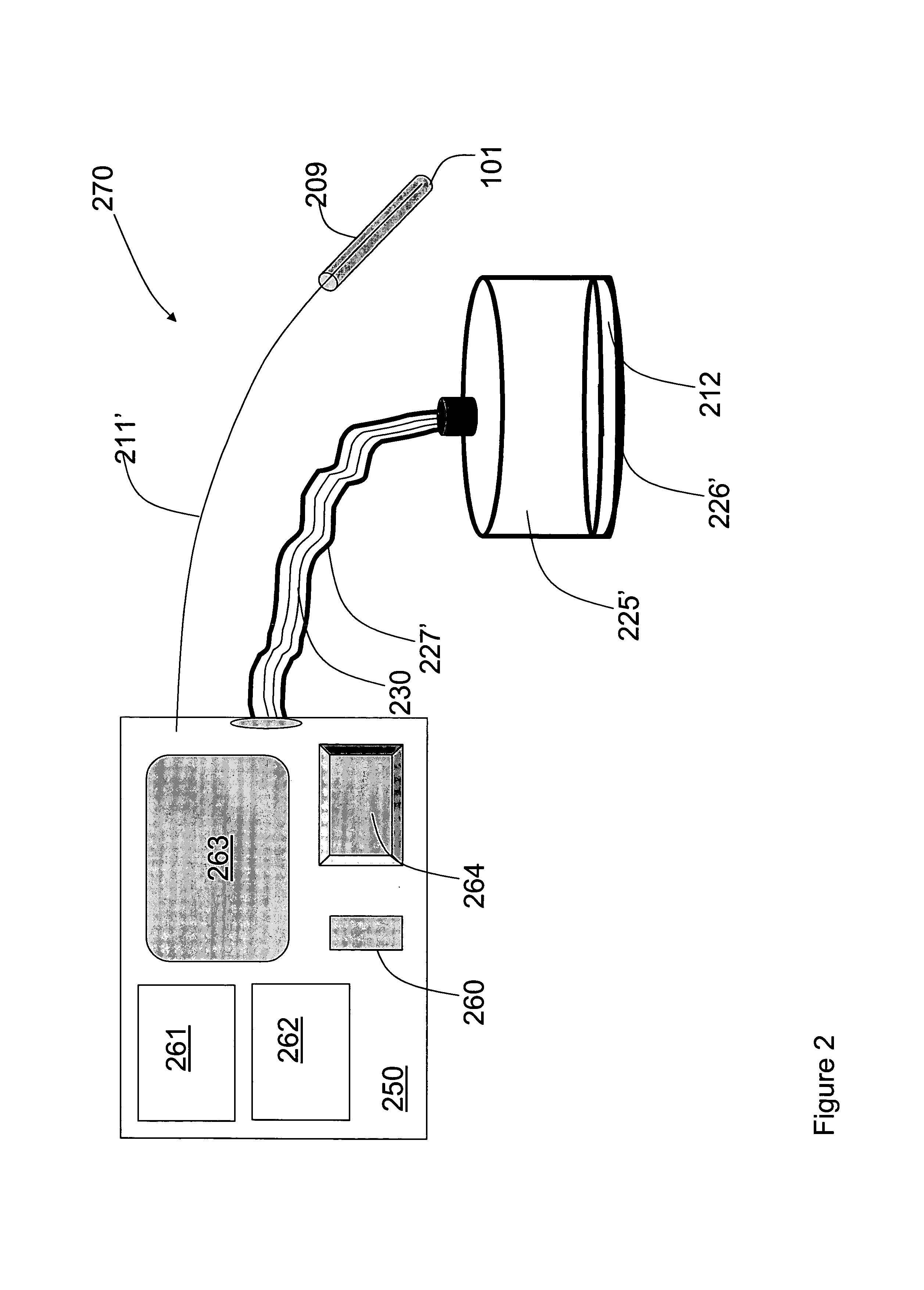
[0055]FIG. 1 shows a schematic diagram of a tissue analyzer 210 of the present invention. The tissue analyzer 210 includes an illumination unit 260 (e.g., a pulsed laser); a processor 261 having a memory 262, a data presentation utility (display) 263 and an input / output control panel 264; and an acoustic unit 212. In the present example, the illumination unit and processor form parts of a common controller 250. A hand held (or fixed) probe head 225 has a probe surface 226 that is adapted for application onto a body surface. The probe head 225 is connected to the controller 250 by light guiding means (optical fiber 211, for example, located in a flexible sheath 227. The optical fiber 211 conducts light energy from the illumination unit 260 to a distal end 101 of the optical fiber 211 located at probe surface 226 in order to irradiate body tissues adjacent to the probe surface 226 when the probe surface is applied to a body surface. The acoustic unit 212 is located in the probe head 2...


PUM

PropertyMeasurementUnit
wavelengthaaaaaaaaaa
timeaaaaaaaaaa

Abstract

A method and apparatus are presented for non-invasively monitoring at least one characteristic of a region of interest in a body. The body is illuminated by pulsed light to induce a photoacoustic effect. Acoustic radiation is detected, and measured data indicative thereof is generated. The photoacoustic effect is controlled by carrying out at least one of the following: providing the pulsed light of at least two different wavelength, analyzing the measured data to determine time variations of at least one predetermined parameter of a time dependent acoustic signal for each of wavelength, and determining oxygen saturation level in the region of interest; and operating a plurality of acoustic elements configured for detection of the acoustic radiation, to thereby define a focal volume for acoustic radiation detection to match dimensions of the region of interest.

Description

RELATED APPLICATIONS[0001]The present application claims the benefit of U.S. Provisional Appln. No. 60 / 502,208, filed Sep. 12, 2003, and U.S. Provisional Appln. No. 60 / 502,209, filed Sep. 12, 2003, the entirety of which are hereby incorporated by reference in their entirety.FIELD OF THE INVENTION[0002]This invention relates to medical devices and methods, and more specifically to such devices and methods for non-invasive analysis of a region of interest in a human body.BACKGROUND OF THE INVENTION[0003]The well being of a mammal (e.g. human) may be assessed and monitored by determining the level of oxygen saturation in the blood or tissue using the method of oximetry or pulse oximetry. Conventional pulse oximetry uses an apparatus that emits light of at least two wavelengths to irradiate a tissue volume and collects light that propagated through a portion of the tissue containing pulsating blood. The collected light at the two wavelengths is analyzed to determine the heart rate of th...

Claims


Application Information

Patent Timeline
Patent Type & Authority Patents(United States)
IPC IPC(8): A61B5/1455G01N
CPCA61B5/0095A61B5/14542A61B5/1464A61B7/00A61B5/02411A61B5/7264
Inventor BALBERG, MICHALSHECHTER, REVITAL PERYOLSHANSKY, MICHAL
Owner ORNIM
Why Eureka
Try Eureka