Pemphigus monoclonal antibody

Inactive Publication Date: 2009-06-23
KEIO UNIV
View PDF1 Cites 12 Cited by
  • Summary
  • Abstract
  • Description
  • Claims
  • Application Information

AI Technical Summary

Benefits of technology

[0008]The present invention relates to: a pemphigus monoclonal antibody having a pathogenic activity that can induce pemphigus lesion (1); a pemphigus monoclonal antibody that specifically binds to human and mouse desmoglein (Dsg) 3 and does not show crossreactivity to human and mouse desmoglein (Dsg) 1 (2); the pemphigus monoclonal antibody according to (1) or (2) above that specifically recognizes multiple particular amino acid residues in an amino acid sequence shown by Seq. ID No. 1 or 2 (3); the pemphigus monoclonal antibody according to (3) above, wherein the multiple particular amino acid residues are at least one of the valine residue at position 3, lysine residue at position 7 and proline residue at position 8, and an aspartic acid residue at position 59 of Seq. ID No. 1 or 2 (4); the pemphigus monoclonal antibody according to (1) to (4) above comprising an H chain having an amino acid sequence shown by Seq. ID No. 4 or an amino acid sequence constructed by adding, deleting or substituting one or more amino acid residue in the amino acid sequence shown by Seq. ID No. 4, and functions as an H chain of anti-human Dsg3 monoclonal Fab antibody, and an L chain having an amino acid sequence shown by Seq. ID No. 6 or an amino acid sequence constructed by adding, deleting or substituting one or more amino acid residue in the amino acid sequence shown by Seq. ID No. 6, and functions as an L chain of anti-human Dsg3 monoclonal Fab antibody (5); and an antibody fragment including an antigen binding site comprised of a variable region of the pemphigus monoclonal antibody according to (1) to (5) above (6).
[0009]Further, the present invention relates to a pemphigus autoimmune disease animal model wherein pemphigus lesion is induced by administration of a hybridoma that produces the pemphigus monoclonal antibody according to (1) to (5) above (7); a method for screening a therapeutic drug for pemphigus autoimmune disease wherein a test substance is administered to the pemphigus autoimmune disease animal model according to (7) above and the degree of lesion of the pemphigus autoimmune disease in the animal model and/or the antibody titer in the sera is evaluated (8); a method for screening a therapeutic drug for pemphigus autoimmune disease wherein the pemphigus monoclonal antibody according to (1) to (5) above o

Problems solved by technology

The production and induction of an autoantibody having a pathogenicity was

Method used


Image

  • Pemphigus monoclonal antibody
  • Pemphigus monoclonal antibody
  • Pemphigus monoclonal antibody

Examples


example a

Method and Material

A-1 (Construction of rDsg3 Antigen Protein and ELISA)

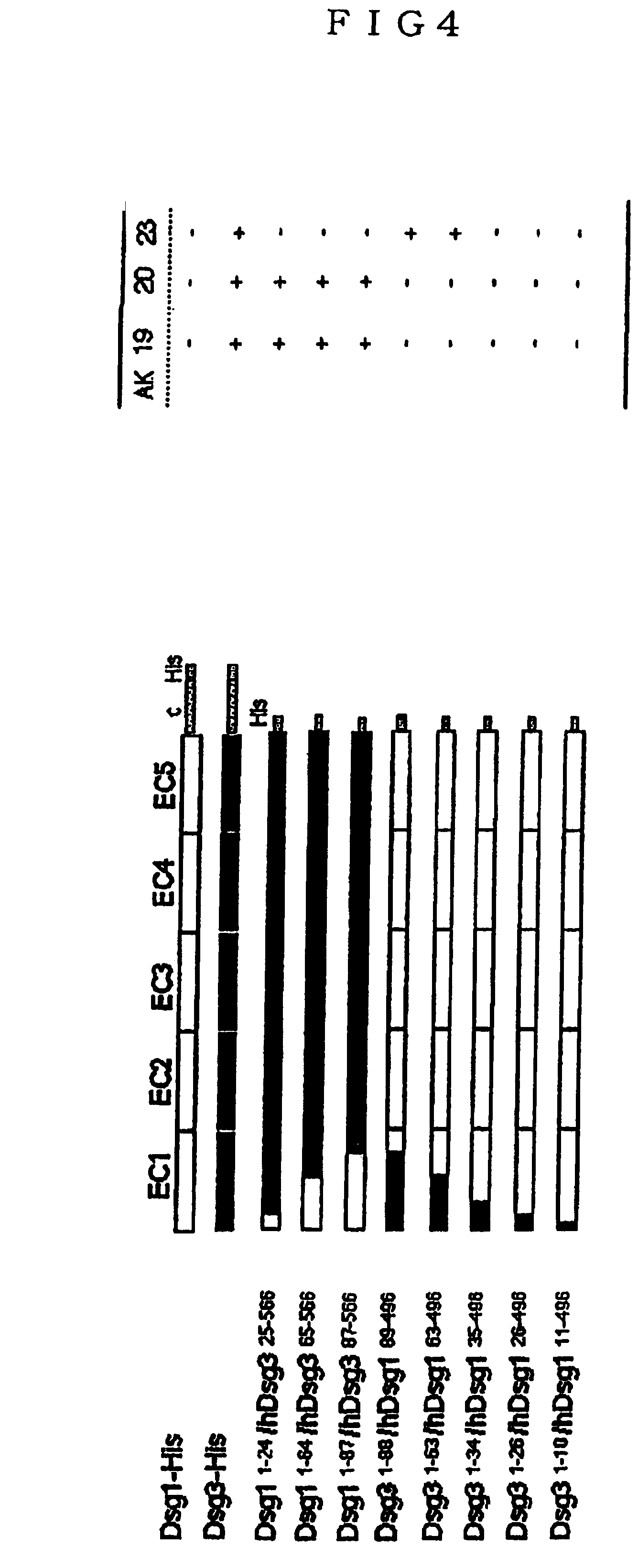
[0028]The baculovirus vectors, pEVmod-Dsg3-His and pEVmod-Dsg1-His, pEVmod-mouse Dsg3-His and pEVmod-mouse Dsg1 -His, that were constructed according to the method as described previously (J. Immunol. 159, 2010-2017, 1997) were used. These recombinant baculoviruses were infected to insect cells, High Five (Invitrogen Corporation), then the recombinant protein human Dsg3-His and Dsg1-His, recombinant protein mouse Dsg3-His and Dsg1-His were collected from the culture media, respectively. In addition, purification of these recombinant proteins was conducted using Ni-NTA column. These purified recombinant proteins were coated on ELISA (Enzyme Linked Immunosorbent Assay) plates, respectively, 50 μl each of the culture supernatant of the anti-Dsg3 monoclonal antibody-producing hybridoma was added to each well, and ELISA was practiced according to the method as previously described (J Clin Invest 105, 625-631, 2000). ...

example b

Results

B-1 (Reactivity of Monoclonal Antibodies)

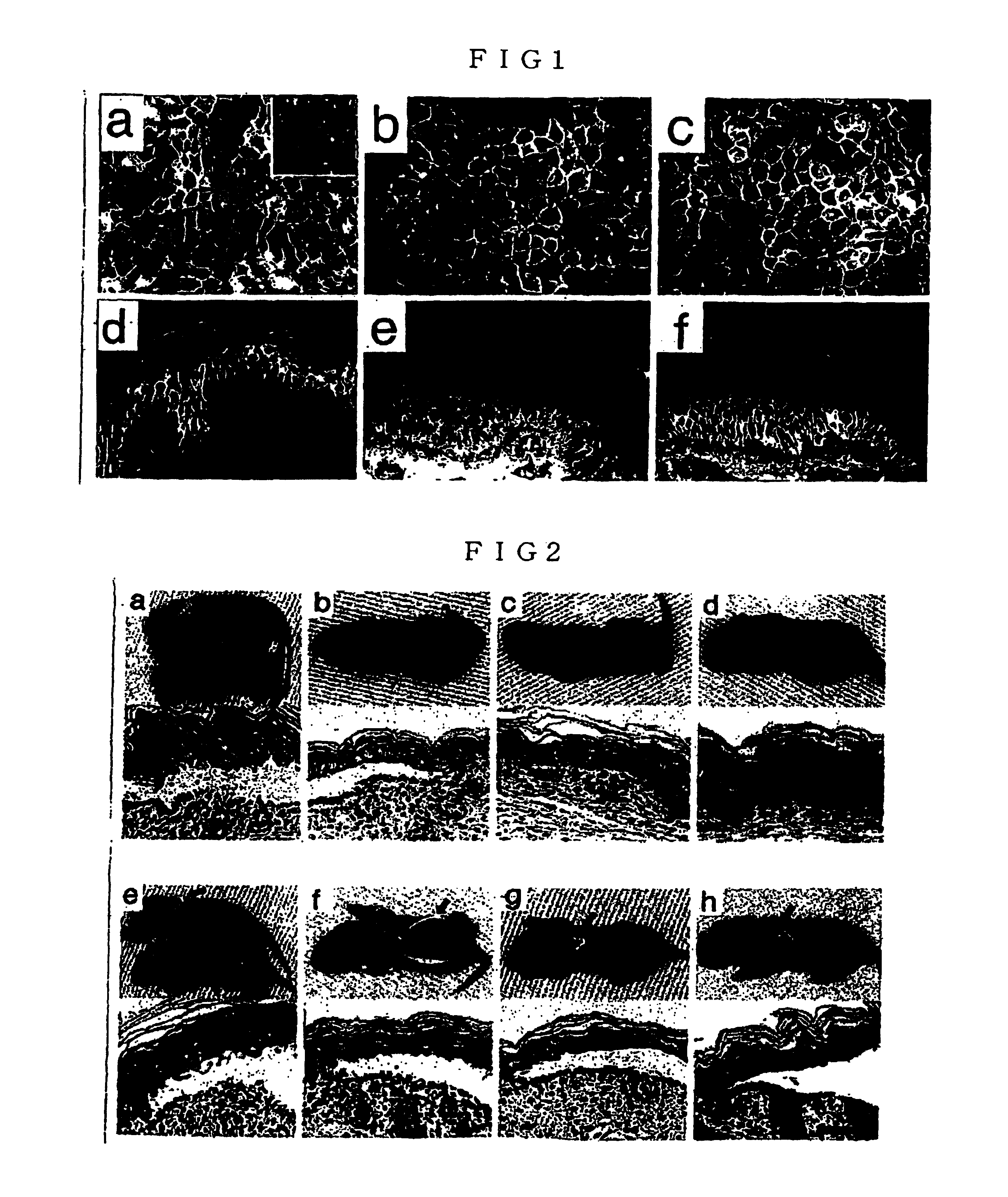
[0044]The PV mouse model with an active disease can induce loss of cell-cell adhesion of keratinocytes, thereby having a circulating anti-Dsg3 IgG antibody that forms blisters. The present inventors used this PV mouse model as a supply source for developing anti-Dsg3 IgG monoclonal antibody. The present inventors produced hybridoma cells from the splenocytes of these mice. First, for these hybridoma cells, recombinant Dsg3 obtained from the baculovirus expression system were used for screening with ELISA, and for positive clones, screening was further performed by living keratinocyte staining using mice keratinocyte PAM212 cells (FIGS. 1a, 1b and 1c). In the screening for the second time, antibodies capable of binding to native Dsg3 at the surface of keratinocytes in vivo were selected, and 8 different clones were isolated (Table 3). The isotypes of these monoclonal antibodies were all IgG1 for H chain and K for L chain.

[0045]ELISA met...


PUM


Abstract

The present invention provides a monoclonal antibody having a pathogenic activity that can induce pemphigus lesion, a peptide specifically recognized by the monoclonal antibody and useful as a therapeutic drug for pemphigus autoimmune disease, etc. As anti-mouse Dsg3 antibody-producing cells are present in the splenocytes of pemphigus vulgaris mouse model constructed by using autoantigen knockout mouse, cell fusion was conducted with the splenocytes of said mouse model and mouse myeloma cells using polyethyleneglycol, hybridomas were constructed, and monoclonal antibodies against Dsg3 were constructed. Among them, a monoclonal antibody having a pathological activity that can induce pemphigus lesions was screened, the base sequence and amino acid sequence in its variable region (heavy chain, light chain) was determined, and the specific epitope part was identified.

Description

TECHNICAL FIELD [0001]The present invention relates to a pemphigus monoclonal antibody having a pathological activity that can induce pemphigus lesions, a peptide specifically recognized by the pemphigus monoclonal antibody that is useful as a therapeutic drug for pemphigus autoimmune disease, etc.BACKGROUND ART [0002]Pemphigus vulgaris (PV) is an autoimmune blistering disease of the skin and mucous membranes, which is histologically characterized by intraepidermal blister formation and immunopathologically characterized by the IgG autoantibody against the keratinocyte cell surface (Stanley, J. R. Pemphigus. In Dermatology in General Medicine. I. M. Freedberg, A. Z. Eisen, K. Wolff, K. F. Austen, L. A. Goldesmith, S. I. Katz, and T. B. Fitzpatrick, eds. McGraw-Hill, New York, 654-666 (1998)). Clinically, patients with pemphigus vulgaris exhibit extensive flaccid blister and erosion. These can occur in every stratified squamous epithelium. Pemphigus vulgaris can be lethal, since fail...

Claims


Application Information

Patent Timeline
IPC IPC(8): C07K14/00C07K17/00A61K38/17A61P31/12A61P37/02A61P43/00C07K16/18C07K16/28C12N5/20C12N15/13G01N33/564
CPCA61K38/177C07K16/18C07K16/28G01N33/564G01N2333/705C07K2317/34A61P17/00A61P31/12A61P37/02A61P37/06A61P43/00
Inventor TSUNODA, KAZUYUKIAMAGAI, MASAYUKINISHIKAWA, TAKEJIKOYASU, SHIGEO
Owner KEIO UNIV
Why Eureka
Try Eureka