System and method for time-of-flight imaging

a time-of-flight imaging and time-of-flight technology, applied in imaging devices, instruments, applications, etc., can solve the problems of increasing the risk of radiation-induced tissue damage and cancer development, the need for increased contrast achieved by using elevated radiation doses, and the potential damage of such doses, so as to facilitate time-of-flight imaging and reduce radiation doses , the effect of reducing x-ray exposur

Active Publication Date: 2010-03-30
MAYO FOUND FOR MEDICAL EDUCATION & RES
View PDF16 Cites 7 Cited by
  • Summary
  • Abstract
  • Description
  • Claims
  • Application Information

AI Technical Summary

Benefits of technology

[0017]The present invention overcomes the aforementioned drawbacks by providing a system and method for producing an x-ray image using a significantly reduced radiation dose. The present invention facilitates time-of-flight imaging (TOFI) of relatively large bodies (e.g., a human body) to provide a set of information from which to reconstruct an image of the body. In particular, the present invention measures the refractive index (i.e., ratio of x-ray velocity through matter as compared to velocity through a vacuum) of x-rays through the body and utilizes this information to reconstruct an image of the body. As such, the present invention is capable of reducing x-ray exposure by orders of magnitude, because the contrast conveyed by the refractive index of x-rays through soft tissues is considerably greater than that obtained with traditional x-ray imaging.

Problems solved by technology

The use of the lack of photons detected as the “signal” from which an image is generated presents a number of inherent problems.
However, high doses of radiation are typically undesirable due to the x-ray's potential to damage the body.
In particular, there is a direct relationship between the number of x-ray photons interacting with tissue and the increased risk of radiation-induced tissue damage and cancer development.
In traditional x-ray imaging systems that rely upon attenuation-based x-ray imaging, a dilemma is created between the need for increased contrast achieved using elevated radiation doses and the potential for damage that such doses can yield.
However, these contrast agents also carry some risks.
Furthermore, enhancement of traditional x-ray imaging is limited to incremental reduction of radiation exposure.
Unfortunately, these attempts for reducing radiation exposure in traditional x-ray imaging systems have been only somewhat successful.
For example, modulating x-ray tube current as a function of beam angle relative to the patient, while first proposed in the mid 1980s, still has not been found to be compelling.
A major problem here is that tuneable monochromatic x-ray sources are still not routinely available for clinical use.
While clearly being theoretically advantageous, velocity-based imaging systems have not been widely realized due to various formidable hurdles to actual implementation.
In particular, while previously demonstrated as operable by imaging small specimens, systems have not been successfully developed that are appropriate for humans or full body imaging.
These approaches are faced, however, with the problem of “unraveling” the many changes in refraction (expressed as phase shifts) that occur as the photon passes through 30 cm or more of tissue.
Hence, for a 30 centimeter (cm) human abdomen, 6000 phase shifts would be expected, which is computationally cumbersome, if not currently impossible, to “unravel”.
However, to implement this approach, a perfect silicon crystal is needed, which is difficult and costly to produce at the large scales necessary to image humans.
In this case, the optic path length of the reference beam should be stable to within 0.1 nanometer (nm), which presents another significant technological impediment to implementation.
However, each of these methods, as well as the others described above, rely on rather low energy x-ray photons (approximately 10-30 keV) having relatively long wave lengths because the low energy x-ray photons yield more obvious interference patterns and greater refraction deviation.
The lower energy x-ray photons are not suitable for imaging larger human subjects because the majority of the photons are stopped by the long tissue path lengths.
Thus, while these methods have been demonstrated as feasible on small specimens, they are not clinically viable for human patients.

Method used


Image

  • System and method for time-of-flight imaging
  • System and method for time-of-flight imaging
  • System and method for time-of-flight imaging

Examples


Embodiment Construction

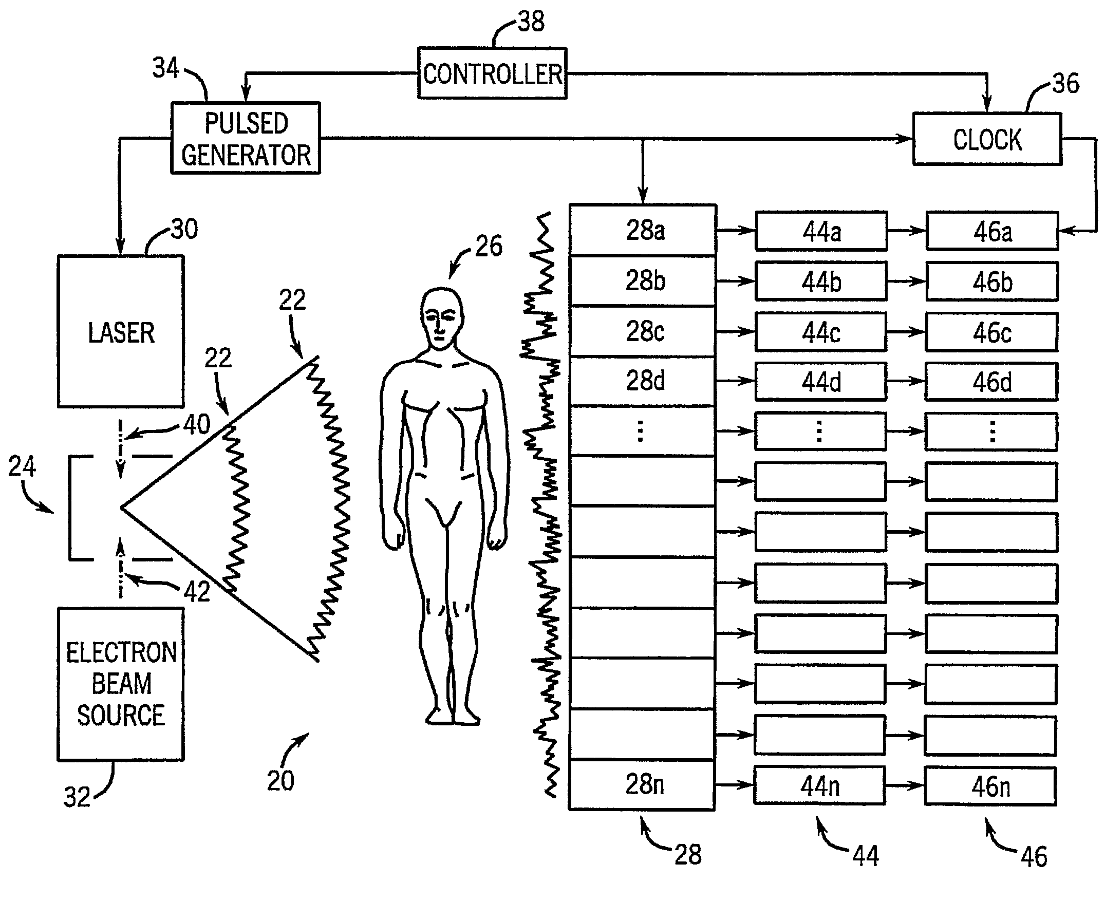
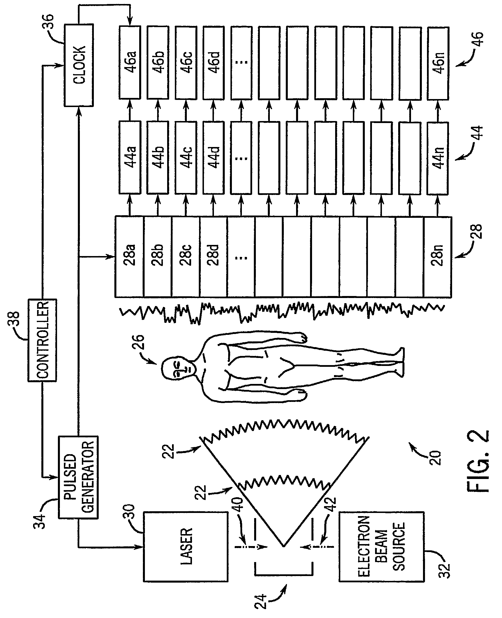
[0032]It is contemplated that a variety of techniques may be employed to perform time-of-flight imaging (TOFI) to image large scale subjects, such as human patients. In particular, three systems usable with two detector systems will be described for clinically applicable imaging of the x-ray refractive index of tissues.

[0033]Referring to FIG. 2, in accordance with one aspect of the invention, one system 20 for performing TOFI is designed to generate a brief x-ray pulse 22 and measure the time of flight from a source 24, through a subject 26, and to a detector system 28. The high intensity x-ray pulse 22 can be generated by aiming a high power laser 30 directly at an oncoming electron beam source 32 to form the source 24. A pulse generator 34 is controlled in conjunction with a clock 36 by a controller 38 to cause the laser 30 to emit periodic pulses of laser light 40. The laser light 40 emitted by the laser 30 collides with and slows down the electrons of an electron beam 42 emitted...


PUM

PropertyMeasurementUnit
optic path lengthaaaaaaaaaa
diameteraaaaaaaaaa
volumeaaaaaaaaaa

Abstract

A system and method for imaging a subject includes a clock that generates a clock signal and a radiation source that directs photons through the subject in response to the clock signal. A detector system is included that detects the photons and a memory module records a time of detection of the photons by the detector system with respect to the clock signal. The system includes a processor that calculates a time of flight (TOF) of the photons from the radiation source to the detector system and compares the TOF to a reference TOF to determine a delay in the TOF attributable to the photons passing trough the subject.

Description

CROSS REFERENCE TO RELATED APPLICATIONS[0001]This application represents the national stage application of International Application PCT / US2006 / 026090, filed 3 Jul. 2006, which is based on U.S. Provisional Patent Application Ser. No. 60 / 696,385 filed on Jul. 1, 2005, and entitled “EXPLORATION OF TIME OF FLIGHT X-RAY IMAGING (TOFXI)”.STATEMENT REGARDING FEDERALLY SPONSORED RESEARCH[0002]N / ABACKGROUND OF THE INVENTION[0003]The present invention relates generally to a system and method for generating high-quality x-ray images using relatively small radiation doses. In particular, the present invention includes a system and method for generating x-ray images by utilizing time-of-flight information to derive contrast information. This time-of-flight information is derived by measuring the refractive index (i.e., ratio of x-ray velocity through matter as compared to velocity through a vacuum) of x-rays through a body, which can then be used to reconstruct an image of the body.[0004]X-ray ...

Claims


Application Information

Patent Timeline
Patent Type & Authority Patents(United States)
IPC IPC(8): G01N23/04H05G1/64
CPCA61B6/00A61B6/484G21K2207/005A61B6/4092
Inventor RITMAN, ERIK LEO
Owner MAYO FOUND FOR MEDICAL EDUCATION & RES
Why Eureka
Try Eureka