
Medical film

a medical film and film technology, applied in the field of medical film, can solve the problems of pain or impair function, difficult follow-up operation with respect to primary disease, and the living body adhesion to one, and achieve the effects of excellent biocompatibility, excellent strength in suturing and bonding

Inactive Publication Date: 2010-05-18
GUNZE LTD
View PDF26 Cites 1283 Cited by
  • Summary
  • Abstract
  • Description
  • Claims
  • Application Information

AI Technical Summary

Benefits of technology

[0007]Therefore, it is an object of the present invention to provide a medical film that is, for example, excellent in biocompatibility and bioabsorbability and has an excellent strength in suturing and bonding.
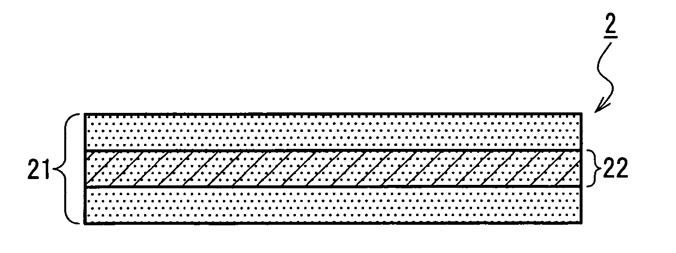
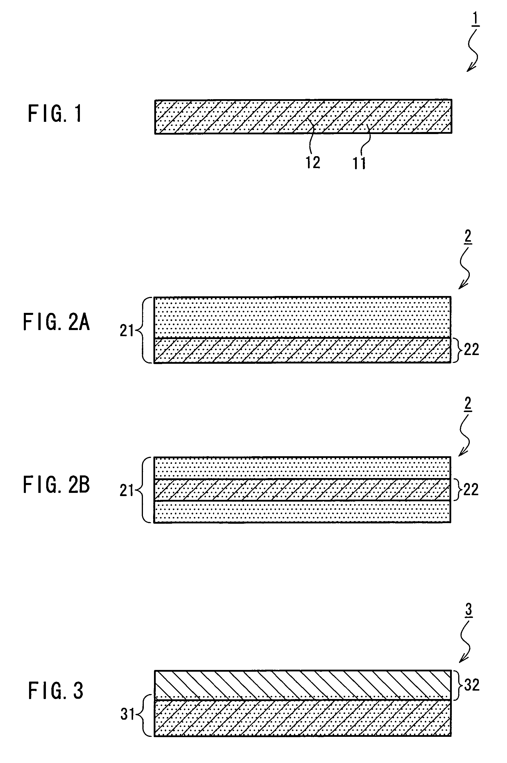
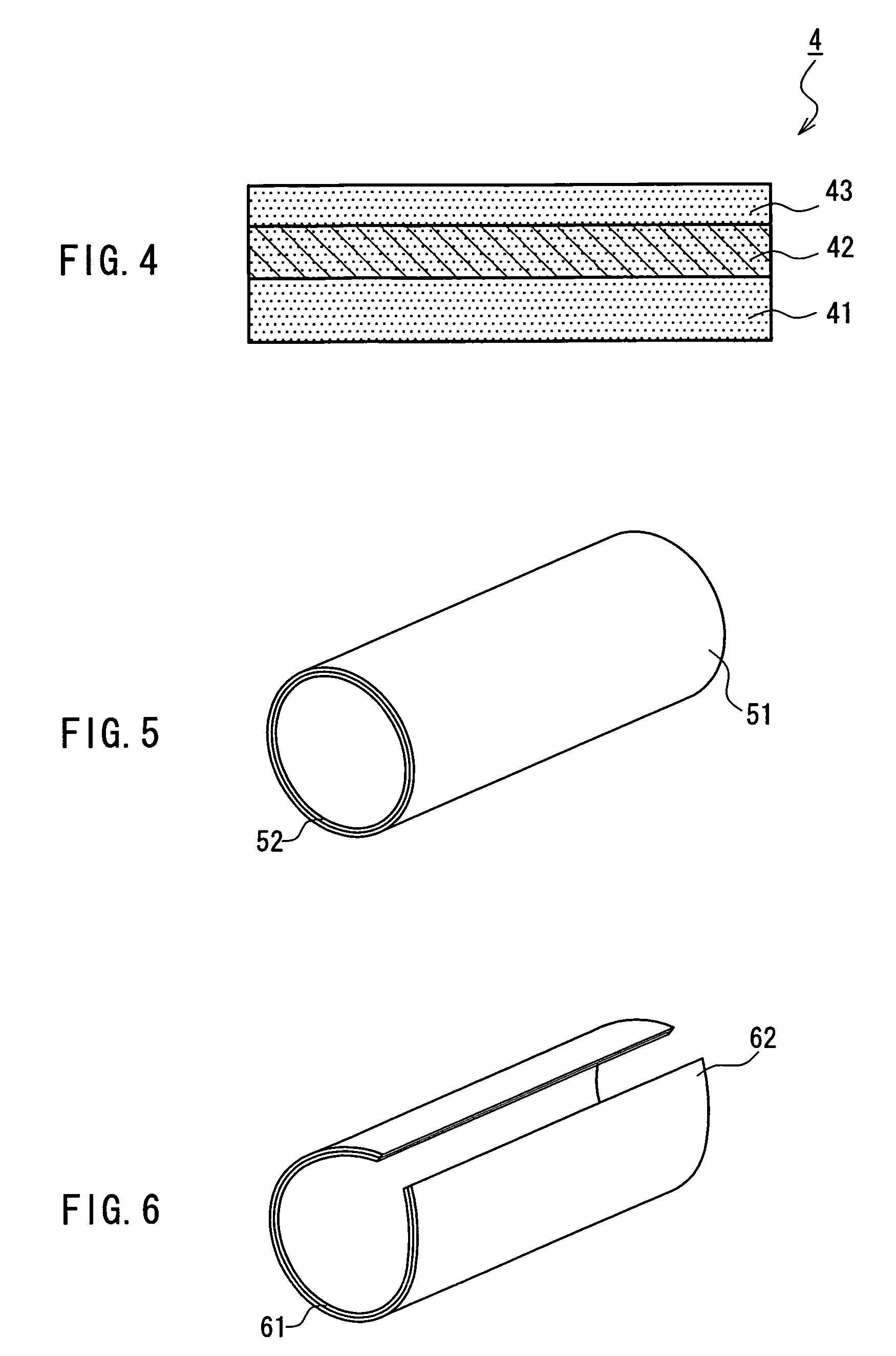
[0009]In the medical film of the present invention, a sufficient strength is imparted by disposing the reinforcing material made of a biodegradable polymer so as to extend over an entire area in a plane direction of at least one of a surface and an internal part of the gelatin film and integrating the reinforcing material with the gelatin film. Therefore, for example, the medical film is easy to fix at an application site, and the fixed state can be maintained during a required period. Accordingly, when the medical film of the present invention is used as, for instance, an antiadhesive material, the adhesion prevention effect due to the gelatin film can be performed sufficiently at the application site. Moreover, since the gelatin film is reinforced in its entirety, there is an advantage that, for example, the gelatin film can be used after being cut into a desired form or a size, so that the application site is not limited. Moreover, for example, even in the case where the medical film that has been fixed at the application site by suturing needs to be peeled off, suturing can be carried out as many times as needed at a portion of the medical film different from the portion that has already been subjected to suturing, because the medical film is reinforced in its entirety. Further, since the reinforcing material is made of a biodegradable polymer with biocompatibility that is field-proven in the clinical medicine, a problem that it remains in a living body and causes a foreign body reaction with tissues, for instance, can be avoided. Therefore, the medical film of the present invention can serve as, for example, an antiadhesive material that is particularly advantageous in the clinical field such as surgical operations.

Problems solved by technology

In various clinical fields including cardiac surgery, orthopedics, neurosurgery, abdominal surgery, and obstetrics and gynecology, it has been a serious problem that after a surgical operation of various types or due to an external injury, tissues in an affected part in a living body adhere to one another.
The adhesion of tissues, for instance, can cause pain or impair function, which, if serious, requires another surgical operation for separating the adhering tissues.
Moreover, the adhesion also causes a problem of making a follow-up operation with respect to the primary disease difficult.
However, the conventional antiadhesive material as described above has difficulty in, for instance, performing the antiadhesive function while maintaining its form in a living body for a required period of time, and since it does not have a strength sufficient for durability in suturing, bonding, or the like, it is torn in some cases.
Thus, the handling of the antiadhesive material and the fixing of the same at an application site are difficult.
For instance, antiadhesive materials formed with gelatin films excellent in biocompatibility, bioabsorbability, etc., which recently have been studied and developed, and now are in actual use (see JP 11(1999)-239610 A and JP 2000-37450 A, for instance) are inferior in allowing themselves to adhere and fix to surfaces of tissues at an application site.
Therefore, they have a problem of being difficult to fix by suturing, etc.

Method used


Image

  • Medical film
  • Medical film
  • Medical film

Examples


example 1

Manufacture of Fabric Body

[0106]Using a lactic acid-caprolactone copolymer multifilament yarn (thickness: 42 decitex (dtex)), a twin loop knit (thickness: 200 μm) in which a vertical length and a horizontal length of a unit of stitches were both 3.5 mm and a twin loop knit (thickness: 200 μm) in which a vertical length and a horizontal length of a unit of stitches were both 1.5 mm were prepared. The “dtex” (decitex: 1 dtex=1.111×1 denier) is a thickness unit according to the International System of Units (SI). As described above, FIG. 7 illustrates a schematic diagram of a knit stitch structure of a twin loop knit. Each of the twin loop knit fabric bodies was held between two glass plates and subjected to a vacuum heat treatment at 120° C. for 3 hours. Subsequently, each of the heat-treated twin loop knit fabric bodies was subjected to a plasma treatment at room temperature, in oxygen gas at 67 Pa (0.5 torr), with 50 W, for 30 seconds.

Integration with Gelatin Film

[0107]Each of the f...

example 2

[0121]As fabric bodies, complex films were prepared using a twin knit and a warp knitted fabric shown below, respectively, and the strength of the thus-obtained films were determined. Note here that a lactic acid-caprolactone copolymer multifilament yarn used for the preparation of the films was prepared using lactic acid-caprolactone copolymer containing lactide (a dimer of lactic acid) and caprolactone at a composition ratio (a molar ratio) of 75:25 by a known method (see JP 8(1996)-317968 A, for example).

Reinforcing Material

[0122](Twin Knit 2-1)

[0123]Using a lactic acid-caprolactone copolymer multifilament yarn (thickness: 75 decitex (dtex)), a twin loop knit in which a vertical length of a unit of stitches was 2.7 mm and a horizontal length of the same was 3.1 mm was prepared. FIG. 10A is a photograph showing a knit stitch structure of the twin knit 2-1 (25× magnification).

[0124](Twin Knit 2-2)

[0125]Using a lactic acid-caprolactone copolymer multifilament yarn (thickness: 67 dec...


PUM

PropertyMeasurementUnit
horizontal lengthaaaaaaaaaa
thicknessaaaaaaaaaa
yarn threading tensionaaaaaaaaaa

Abstract

A medical film that is excellent in biocompatibility and bioabsorbability and has an excellent strength in suturing and bonding is provided. A reinforcing material 12 made of a biodegradable polymer is placed in a gelatin solution so as to allow the solution to infiltrate in the reinforcing material 12 and then the gelatin is dried. This allows the gelatin that has infiltrated entirely in an internal part of the reinforcing material 12 to gel, thereby forming a gelatin film 11. Thus, a medical film 1 in which the reinforcing material 12 and the gelatin film 11 are integrated is obtained. The gelatin film 11 preferably is a cross-linked gelatin film.

Description

TECHNICAL FIELD[0001]The present invention relates to a medical film. In particular, the present invention relates to an antiadhesive material for preventing tissues in a living body from adhering to each other, a tissue prosthetic material for prosthetic restoration of a missing part of a tissue, or a graft cell-culturing sheet material for implanting a sheet-like tissue in a living body, which are excellent in biocompatibility and bioabsorbability and have an excellent strength in suturing.BACKGROUND ART[0002]In various clinical fields including cardiac surgery, orthopedics, neurosurgery, abdominal surgery, and obstetrics and gynecology, it has been a serious problem that after a surgical operation of various types or due to an external injury, tissues in an affected part in a living body adhere to one another. The adhesion of tissues, for instance, can cause pain or impair function, which, if serious, requires another surgical operation for separating the adhering tissues. Moreov...

Claims


Application Information

Patent Timeline
Patent Type & Authority Patents(United States)
IPC IPC(8): A61F2/02A61F2/06B32B27/04B32B27/12B32B5/04A61L31/04A61L31/12A61L31/14
CPCA61L31/045A61L31/148A61L31/129Y10T442/2484Y10T442/2525Y10T442/2861Y10T442/3854Y10T442/45Y10T442/674
Inventor MATSUDA, SHOJIROOHTANI, HITOSHITANAKA, YOSHIMITADOKORO, HIDEKI
Owner GUNZE LTD
Learn More