Cell growth inhibitors containing anti-glypican 3 antibody
a cell growth inhibitor and anti-glypican technology, which is applied in the field of cell growth inhibitors containing antiglypican 3 antibodies, can solve the problems of no clear relationship between
- Summary
- Abstract
- Description
- Claims
- Application Information
AI Technical Summary
Benefits of technology
Problems solved by technology
Method used
Image
Examples
example 1
Preparation of Monoclonal Antibody Against Glypican-3 Synthetic Peptide
[0093]A peptide having an amino acid sequence (the 355th to the 371st amino acids) (Arg Gln Tyr Arg Ser Ala Tyr Pro Glu Asp Leu Phe Ile Asp Lys Lys of human glypican-3 protein (SEQ. ID NO. 1) was synthesized. The synthetic peptide was bound to keyhole limpet hemocyanin (KLH) using a maleinimide benzoyloxy succinimide (MBS) type crosslinking agent, thereby preparing an immunogen. Mice (BALB / c, female, 6-week-old) were immunized 3 times with the immunogen at 100 μg / mouse. The antibody titers in serum were assayed. A method employed as an antibody titer assay method involves causing the diluted sera to react with the peptides (0.5 μg) immobilized on a plate, performing reaction with HRP-labeled anti-mouse antibodies, adding a substrate, and then measuring absorbance at 450 nm of the thus developed color (a peptide solid-phase ELISA method). After antibody titers were confirmed, splenocytes were collected, and then f...
example 2
Inhibition of Cell Proliferation Using Anti-Glypican 3 Antibody
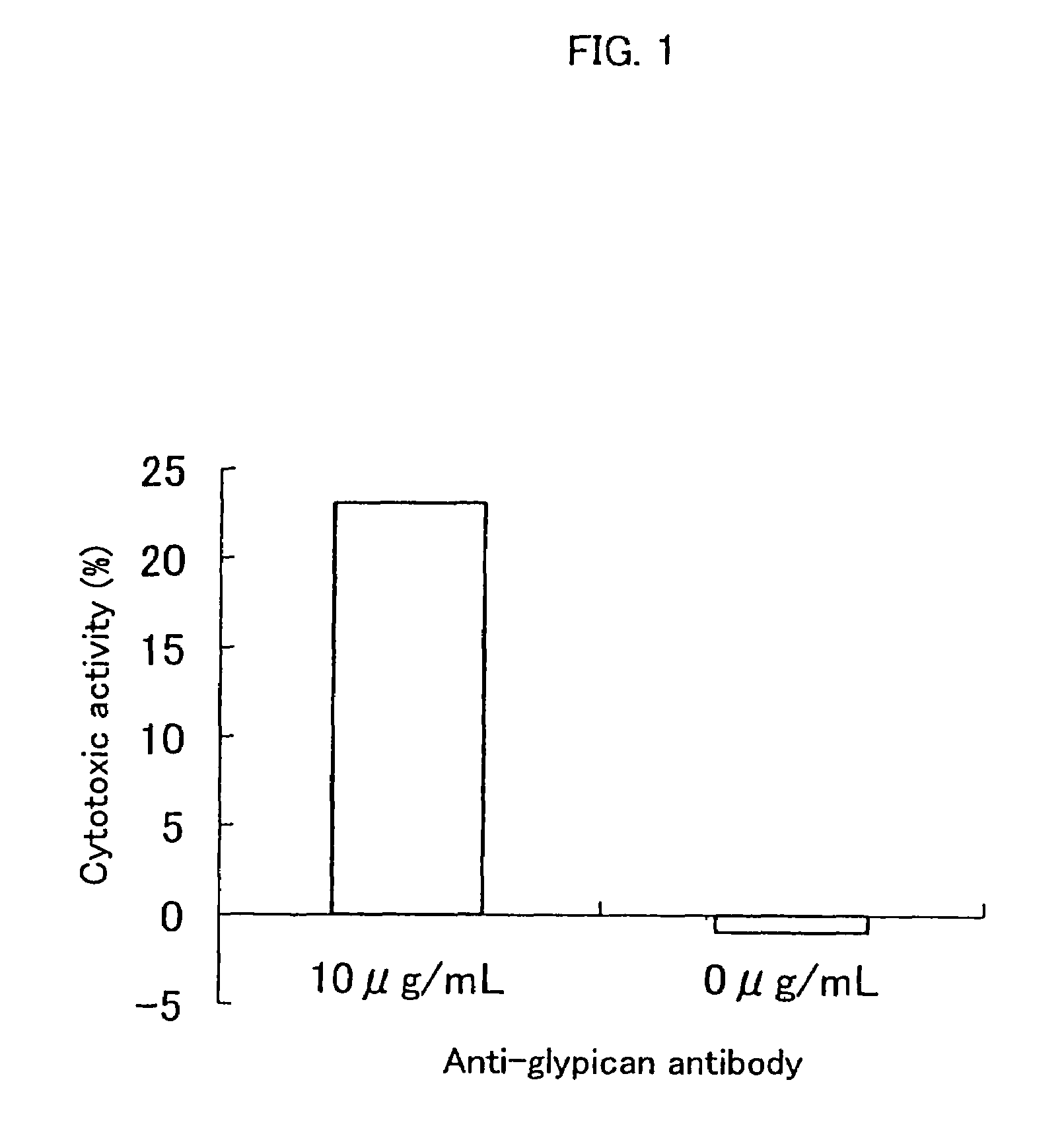
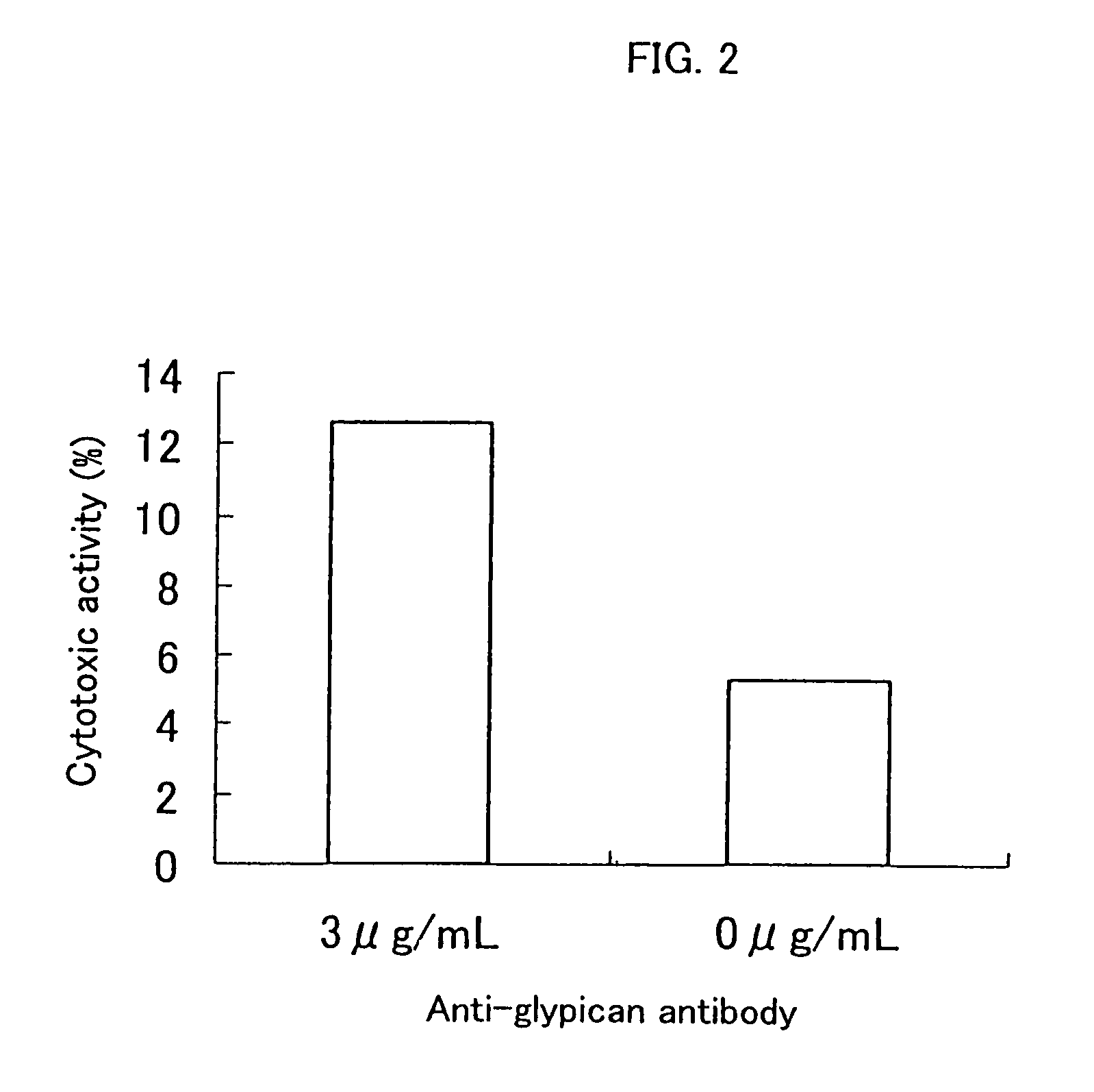
[0094]The ADCC (antibody-dependent cell-mediated cytotoxicity) activity and the CDC (complement-dependent cytotoxicity) activity were measured according to the method of Current Protocols in Immunology, Chapter 7. Immunologic studies in humans, Editor, John E, Cologan et al., John Wiley & Sons, Inc., 1993.
1. Preparation of Effector Cell
[0095]The spleen was extracted from a CBA / N mouse (8-week-old, male), and then splenocytes were isolated in RPMI1640 media (GIBCO). The cells were washed in the same media containing 10% fetal bovine serum (FBS, HyClone), and then the cell concentration was prepared at 5×10⁶ / mL, thereby preparing effector cells.
2. Preparation of Complement Solution
[0096]Baby Rabbit Complement (CEDARLANE) was diluted 10 times in 10% FBS-containing DMEM media (GIBCO), thereby preparing a complement solution.
3. Preparation of Target Cell
[0097]An HuH-7 human hepatic cancer cell line (Japanese Collection of Res...
example 3
Measurement of the Expression Level of Glypican on HuH-7 Cells
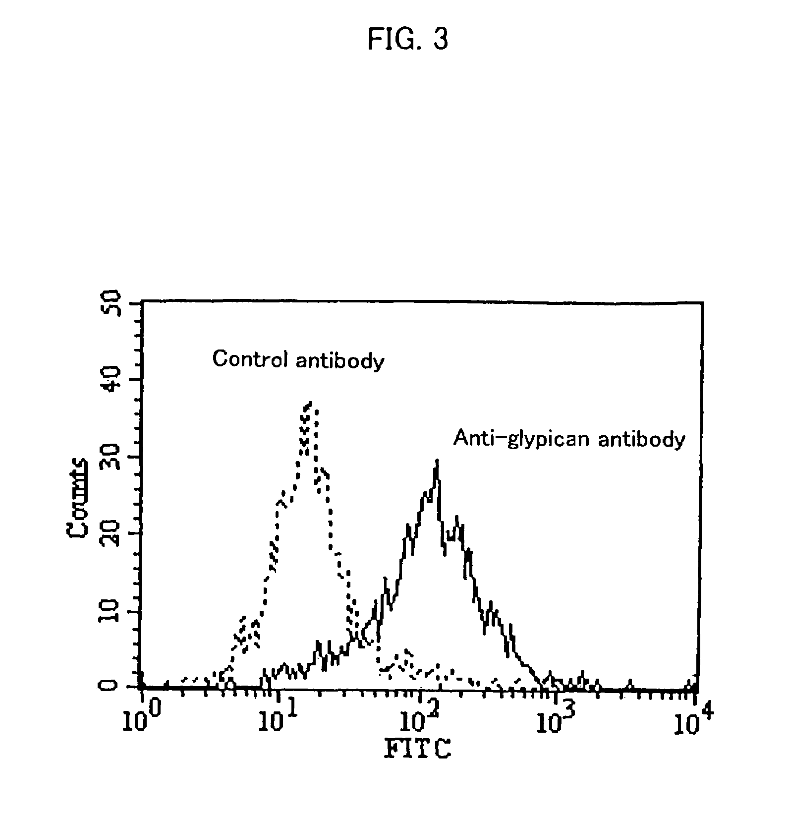
[0101]Approximately 5×10⁵ HuH-7 cells were suspended in 100 μL of FACS / PBS (prepared by dissolving 1 g of bovine serum albumin (SIGMA) in 1 L of CellWASH (Beckton Dickinson)). Then, anti-glypican 3 antibodies (K6511) or mouse IgG2a (Biogenesis) as a control antibody were added at 25 μg / mL, and then the solution was allowed to stand on ice for 30 minutes. After washing with FACS / PBS, the product was suspended in 100 μL of FACS / PBS. 4 μL of Goat Anti-Mouse Ig FITC (Becton Dickinson) was added, and then the solution was allowed to stand on ice for 30 minutes.
[0102]After washing twice with FACS / PBS, the product was suspended in 1 mL of FACS / PBS. Fluorescence intensity of the cells was measured using a flow cytometer (EPICS XL, BECKMAN COULTER).
[0103]FIG. 3 shows the result of flow cytometry. Glypican 3 was expressed on HuH-7 cells, suggesting that anti-glypican 3 antibodies bind to glypican 3 expressed on the cell so as to in...
PUM
| Property | Measurement | Unit |
|---|---|---|
| molecular weight | aaaaa | aaaaa |
| concentration | aaaaa | aaaaa |
| concentration | aaaaa | aaaaa |
Abstract
Description
Claims
Application Information
 - R&D
- Intellectual Property
- Life Sciences
- Materials
- Tech Scout
- Unparalleled Data Quality
- Higher Quality Content
- 60% Fewer Hallucinations
Browse by: Latest US Patents, China's latest patents, Technical Efficacy Thesaurus, Application Domain, Technology Topic, Popular Technical Reports.
© 2025 PatSnap. All rights reserved.Legal|Privacy policy|Modern Slavery Act Transparency Statement|Sitemap|About US| Contact US: help@patsnap.com


