Monoclonal antibody which binds cMet (HGFR) in formalin-fixed and paraffin-embedded tissues and related methods

a monoclonal antibody and cmet technology, applied in the field of molecular diagnostics and medicine, can solve the problems of unreliable commercially available met antibodies, unreliability of immunohistochemical studies, and elevated hgf/sf concentrations in the tumor microenvironment, and achieve the effects of improving immunohistochemical results and improving immunohistochemical results

Inactive Publication Date: 2011-02-22
VAN ANDEL RES INST +1
View PDF2 Cites 28 Cited by
  • Summary
  • Abstract
  • Description
  • Claims
  • Application Information

AI Technical Summary

Problems solved by technology

However, the reliability of the immunohistochemical studies has been questioned in light of the lot-to-lot variability of the antiserum raised against the C-terminal peptide in the c-Met receptor, that has been used in most studies26.
Most commercially available Met antibodies are unreliable (or have not been stringently tested for their reliability).
In addition to increased c-Met expression, elevated HGF / SF concentrations in the tumor microenvironment have also been associated with adverse outcome.
However, the molecular diagnostic tools to identify cancers that possess active Met pathways are not available.
Drugs that bind cell surface receptors or permeate into cells and inhibit receptor and non-receptor kinases show immense promise in the clinic, however the detection of the corresponding targets in human cancers provides a major challenge.
Besides the Hercept test for quantitative measurement of Her-2 / Neu expression, which required a lengthy and arduous development for use in routine clinical samples and FDA approvement, no validated diagnostic tests for receptor or non-receptor protein kinase expression are available.
The difficulty in developing these diagnostic reagents stems from the low level expression of kinases, the labile activation state, which depends on protein phosphorylation and the poor specificity of antibodies against most phospho-epitopes, which indicate kinase activity.
Consequently, most receptor tyrosine kinases (RTK) lack detection reagents for expression measurements in formalin-fixed paraffin embedded (FFPE) tissues, which is the most commonly obtained tissue preparation from patient cancers.
Thus, if patients are not carefully selected for treatment, many agents could fail to demonstrate efficacy in phase II and phase III clinical trials.
The c-Met receptor is particularly difficult to measure in FFPE tissues because of poor reagent choices, insufficient validation of the performance of c-Met antibodies in FFPE tissues and sensitivity of c-Met to formalin-fixation.

Method used


Image

  • Monoclonal antibody which binds cMet (HGFR) in formalin-fixed and paraffin-embedded tissues and related methods
  • Monoclonal antibody which binds cMet (HGFR) in formalin-fixed and paraffin-embedded tissues and related methods
  • Monoclonal antibody which binds cMet (HGFR) in formalin-fixed and paraffin-embedded tissues and related methods

Examples


example 1

Materials and Methods for Examples 1-5

[0079]Monoclonal Antibody Generation and Validation:

[0080]Recombinant protein Met 25-567H was prepared by Dr. Eric Xu's lab in Van Andel Institute. Met 25-567H construct (SEQ ID NO. 1, amino acids at positions 25-567) and purified Met 928 protein were from Dr. Ermanno Gheradi's lab in Cambridge Antibody Technology41. Recombinant fusion protein Met-IgG was purchased from R&D systems (Minneapolis, Minn.).

[0081]Mouse monoclonal antibodies against Met were produced by injecting BALB / c mice i.p. with native and denatured (boiling in SDS sample buffer) Met 25-567H in complete Freund's adjuvant, followed by two additional injections with incomplete Freund's adjuvant. After 1 month, a final injection was given i.p. and i.v. without adjuvant. Polyclonal antisera from immunized mice were tested by indirect immunofluorescence with formalin-fixed MKN45 (Met positive) and NIH3T3 (Met negative) cells. Spleen cells were fused with P3X63AF8 / 653 myeloma cells us...

example 2

Development of an Anti-c-Met Monoclonal Antibody for Quantification of c-Met in Formalin-Fixed Tissues

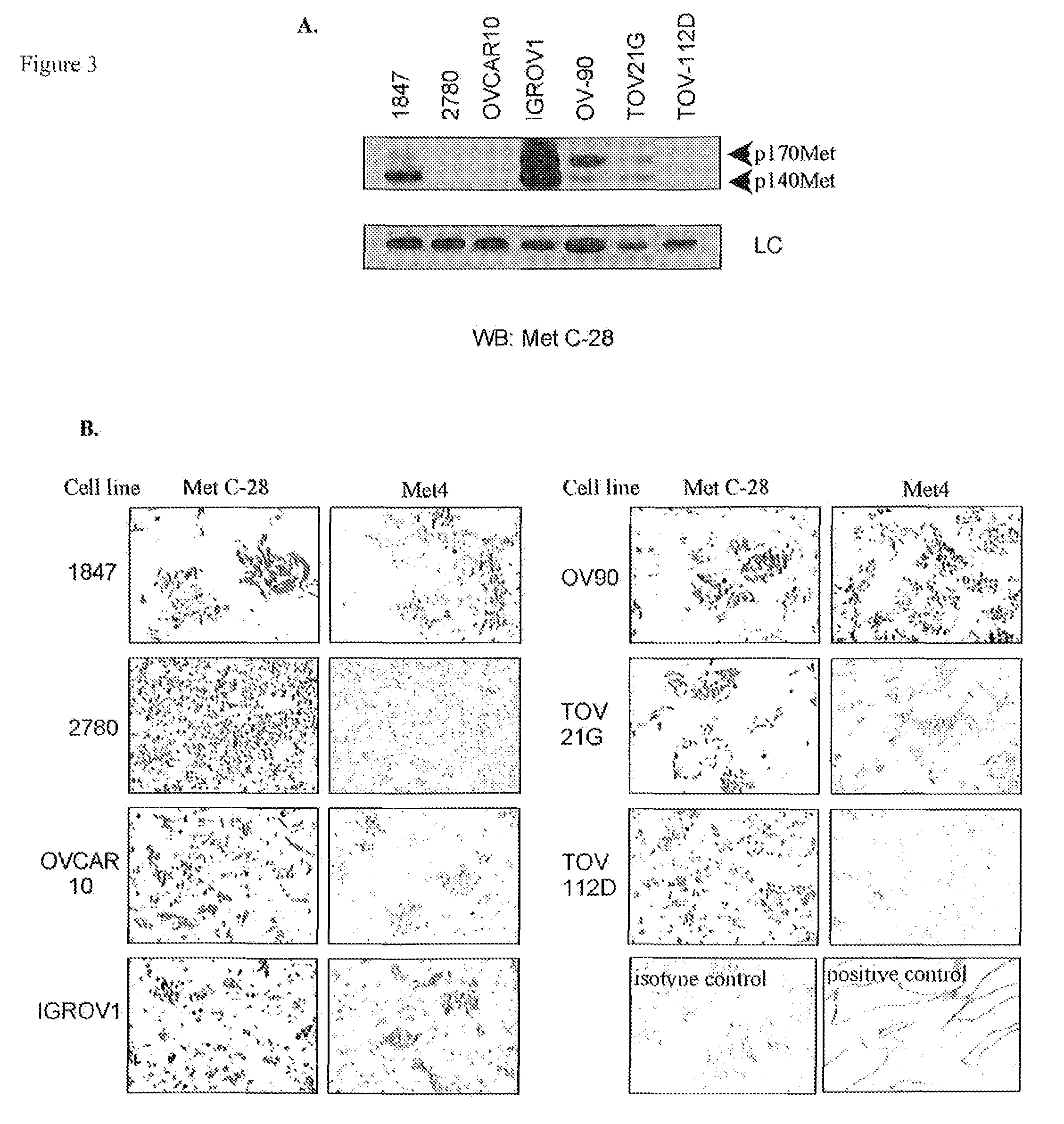
[0095]The limitations of C-28, a polyclonal antibody from Santa Cruz, which has been used to measure Met expression in most in vitro and immunohistochemical studies, motivated our development of a monoclonal antibody (mAb) for measurement of c-Met protein expression in patient cancers for clinical applications, including molecular imaging and immunohistochemistry in formalin-fixed tissues. The immunogen was designed to obtain c-Met antibodies that react with the extracellular domain of the Met receptor, since this provides the most direct measurement to evaluate the abundance of c-Met expression on the cell surface. The resulting mAbs overcome the lot-to-lot variability of C-28 and the nuclear reactivity observed with antibodies that bind the Met cytoplasmic domain.

[0096]As a final screen to identify monoclonal antibodies that specifically bind the Met receptor in formalin-fixed and...

example 3

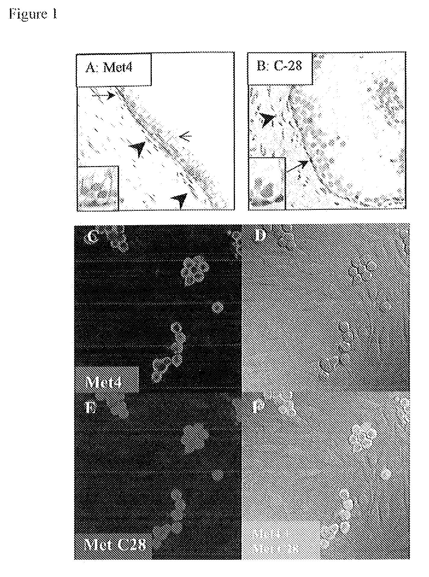
Specificity of Met4 for Reactivity with c-Met in Formalin-Fixed Cells

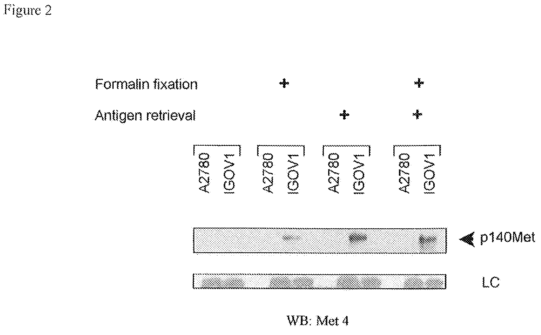
[0099]To confirm the reactivity of Met4 with the Met receptor protein, a Western blot was probed with Met4. By recapitulating conditions of formalin-fixation in tissues, Western blot membranes were treated with 10% formalin and underwent the same type of antigen retrieval as had been applied to tissue sections. Met4 did not react with denatured Met receptor protein in IGROV1 ovarian cancer cells, which express a large amount of c-Met. Formalin fixation or boiling of the membrane in antigen retrieval buffer increased Met4 binding to c-Met and revealed a 140 kDa c-Met band (FIG. 2). Met4 reacted nonspecifically with lower molecular weight bands, some of which also appeared in the Met receptor negative A2780 cell line. The immunofluorescence and Western blotting results demonstrate that Met4 binds to a specific epitope on Met that is sensitive to denaturation and that is re-established by formalin-fixation or antigen ...


PUM


Abstract

In a wide variety of human solid tumors, an aggressive, metastatic phenotype and poor clinical prognosis are associated with expression of the receptor tyrosine kinase Met. Disclosed herein are (a) a monoclonal antibody named Met4, which antibody is specific for Met, and (b) a hybridoma cell line that produces Met4. The Met4 antibody is particularly useful for detecting Met in formalin-fixed tissue. Methods of using the Met4 antibody for detection, diagnosis, prognosis, and evaluating therapeutic efficacy are provided.

Description

FIELD OF THE INVENTION[0001]The present invention is in the field of molecular diagnostics and medicine.BACKGROUND OF THE INVENTION[0002]The c-Met receptor kinase regulates cellular proliferation, migration, differentiation and branching morphogenesis during development and homeostasis1-3. Met is also expressed on the cell surface of a variety of human primary solid tumors and in their metastases (www.vai.org / met / ). The amino acid sequence of the extracellular domain of human Met is provided in SEQ ID NO:1, amino acids 25-567. In its activated state, the Met receptor controls growth, invasion, and metastasis of cancer cells through multiple signal transduction pathways4. In some cancer cell lines, loss of Met expression through silencing promotes apoptosis, demonstrating that Met is necessary for survival5-7. Met activity increases through mutations in the kinase or juxtamembrane domains8-11, through overexpression12, 13, or through binding to its ligand, hepatocyte growth factor (H...

Claims


Application Information

Patent Timeline
Patent Type & Authority Patents(United States)
IPC IPC(8): C07K16/30G01N33/554G01N33/563G01N33/574G01N33/577C12N5/20C12P21/08G01N33/543
CPCC07K16/2863Y10S435/975C07K2317/34
Inventor CAO BRIAN, BOLIANGVANDE WOUDE, GEORGE F.KNUDSEN, BEATRICE S.
Owner VAN ANDEL RES INST
Why Eureka
Try Eureka