Method for magnetic resonance imaging using inversion recovery with on-resonant water suppression including MRI systems and software embodying same
a magnetic resonance imaging and inversion recovery technology, applied in the field of methods, to achieve the effect of increasing the contrast-to-noise ratio and specificity of the magnetic susceptibility gradient, and enhancing the associated signal
- Summary
- Abstract
- Description
- Claims
- Application Information
AI Technical Summary
Benefits of technology
Problems solved by technology
Method used
Image
Examples
example 1
[0109]This example concerns imaging of vascular devices. A saline phantom containing a drug-eluting stainless steel stent (SS), an undeployed MR-compatible nitinol stents (Nitinol), and an MR-compatible imaging guidewire (MRIG) were imaged on a 1.5 T scanner using the IRON sequence. Two-dimensional (2D) slices and 3-dimensional (3D) volumes were acquired with and without the double inversion recovery fat suppression RF pulses. Images using the suppression based imaging methods of the present invention were acquired using fast spin echo (FSE) imaging and the imaging parameters were 180 mm field of view (FOV), 2000 ms repetition time (TR), 7 ms echo time (TE), 0.7×0.7 mm in-plane resolution (RES), 2 signal averages (NSA), and 24 echo train length (ETL). There are shown in FIG. 7A-C various cross-sectional images of the devices placed on a bottle containing copper sulfate.
[0110]In FIG. 7A, the image was acquired without fat suppression and the bandwidth of the spectrally-selective RF p...
example 2
[0112]This example concerned imaging of vascular devices in real time. In this example, the suppression methods of the present invention were used in conjunction with a Gradient and Spin Echo (GRASE) image acquisition technique [Oshio, K. and D. A. Feinberg. GRASE (gradient- and spin-echo) imaging: a novel fast MRI technique. Magn Reson Med 1991; 20 (2): 344-349]. The saline phantom and devices were imaged using an interactive GRASE sequence (180 mm FOV, 158 ms acquisition time(TA); 5.8 ms TE; 4×4×5 mm3; and turbo factor of 11) while the phantom was moved in the magnet. A single cross-sectional image of the phantom is shown in FIG. 9A with low resolution images of the nitinol and stainless steel stents. The trajectory in the x direction of the stainless steel stent (red arrows) over time (t) is shown in FIG. 9B.
example 3
[0113]This example concerns imaging of endovascular devices in vivo.
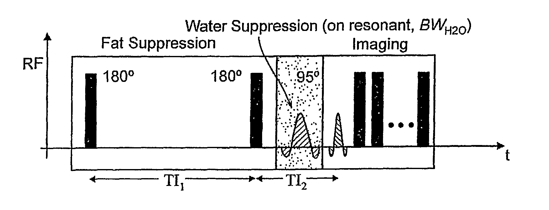
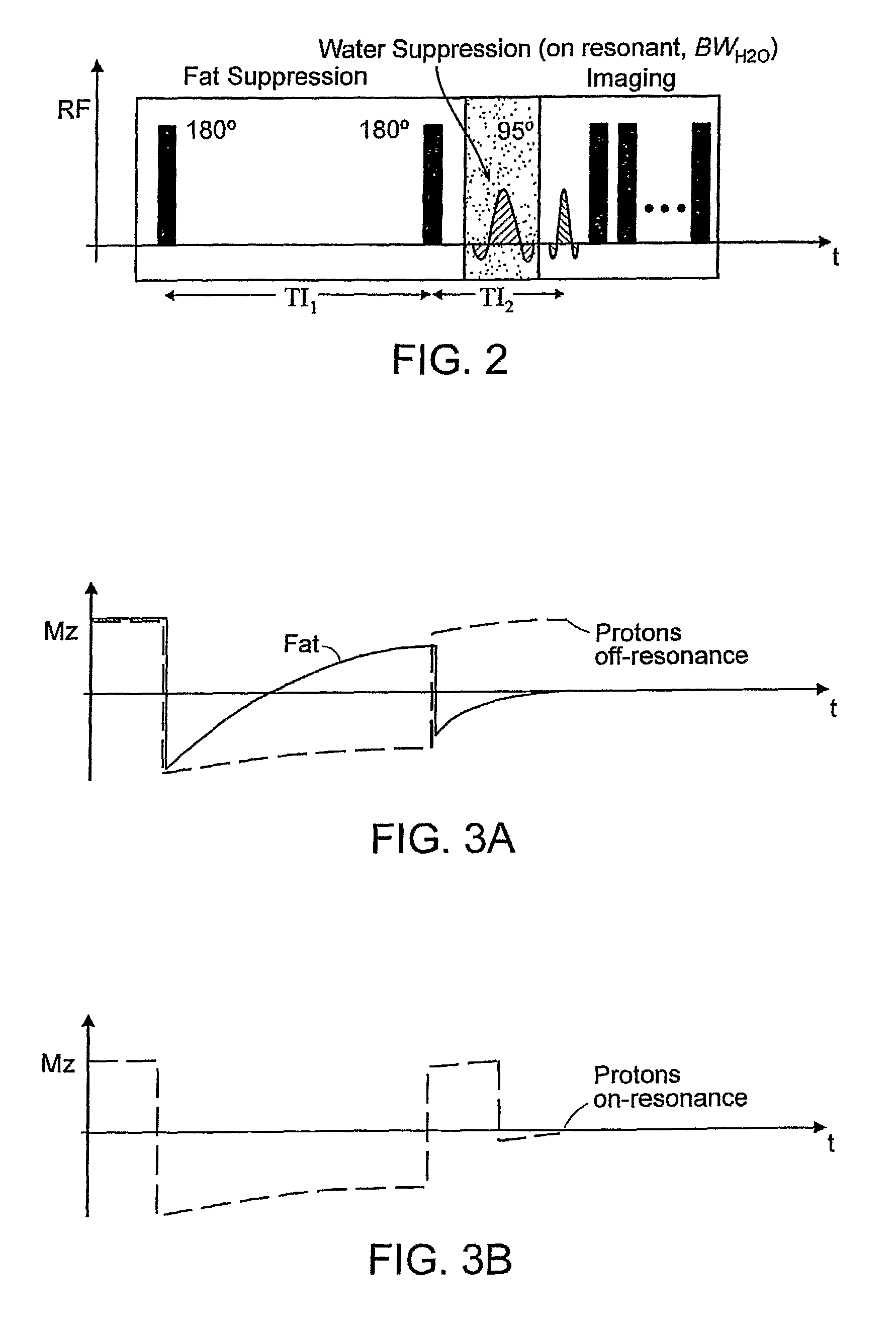
[0114]A rabbit model of hind limb ischemia was employed, whereby platinum coils were placed percutaneously into the left femoral artery under X-ray—fluoroscopy. Two- and Three-dimensional MR images according to the methodology of the present invention were acquired using FSE imaging and the imaging parameters were 180 mm FOV, 2000 ms TR, 8.6 ms TE, 0.35×0.35×1.5 mm3 RES, 4 NSA, and 24 ETL. The excitation radio frequency (RF) pulse for fat suppression had a flip angle (αfat) of 95 degrees and duration (τfat) of 15 ms, and the excitation RF pulse for water suppression had a flip angle (αwater) of 90 degrees and duration (τwater) of 35 ms. GRE imaging of the same slices was performed with 270 mm FOV, 13.6 ms TR, 3.7 ms TE, 20 degree FA, 0.35×0.35×1.5 mm3 RES, 2 NSA, and 12 ETL. GRE and MRI images according to the present invention of the platinum coils in the left femoral artery of a rabbit are shown in FIGS. 10A,B. In...
PUM
 Abstract
Description
Claims
Application Information
 - R&D
- Intellectual Property
- Life Sciences
- Materials
- Tech Scout
- Unparalleled Data Quality
- Higher Quality Content
- 60% Fewer Hallucinations
Browse by: Latest US Patents, China's latest patents, Technical Efficacy Thesaurus, Application Domain, Technology Topic, Popular Technical Reports.
© 2025 PatSnap. All rights reserved.Legal|Privacy policy|Modern Slavery Act Transparency Statement|Sitemap|About US| Contact US: help@patsnap.com


