Method for detecting misfolded proteins and prions

a protein and protein technology, applied in the field of protein detection in biological samples, can solve the problems of protein loss activity, abnormal behavior, formation of high molecular weight deposits or plaques within affected cells, etc., and achieve the effect of increasing detection sensitivity, superior sensitivity and effective detection

Active Publication Date: 2013-02-12
PRESYMPTO INC
View PDF64 Cites 5 Cited by
  • Summary
  • Abstract
  • Description
  • Claims
  • Application Information

AI Technical Summary

Benefits of technology

[0014]The present invention addresses needs in the art, including increasing the detection sensitivity, by providing methods and kits for detection of conformationally-altered proteins, such as prions, which are associated with any number of neurodegenerative diseases and disorders. Conformationally dependent peptides, such as those that are labeled peptides designed to mimic the folding reaction of PrPC converting to PrPSc (also known as, and used equivalently for, PrPTSE), provide the basis for a highly sensitive diagnostic assay for misfolded proteins, such as PrPSc. Superior sensitivity is achieved due to signal amplification as additional peptides, such as labeled peptides, are recruited in a target-enriched solution to undergo a similar conformational change, enabling the effective detection of the misfolded protein, such as PrPSc, in the blood or other tissues of animals and humans containing the misfolded proteins, such as those infected with certain PrPSc-related diseases or those with Alzheimer's disease.

Problems solved by technology

Misfolding of naturally occurring normal cellular proteins is thought to cause the protein to lose activity, or, in many cases, behave abnormally.
Misfolding of normal cellular proteins often causes formation of high molecular weight deposits or plaques within affected cells.
Indeed, most of the diseases and disorders are neurodegenerative in nature, causing diminished neural cell function or neural cell death.
However, prions possess an innate capacity to convert their structure and cause several deadly brain diseases of the dementia type in humans and animals.
Cycling of the protein from the surface to the interior is known to occur, but the function of this cycling is not yet elucidated.
Similarly, by the time post mortem analysis of animal tissue has been completed, products from the animal may have already entered the food stream and potentially infected other animals or even humans.

Method used


Image

  • Method for detecting misfolded proteins and prions
  • Method for detecting misfolded proteins and prions
  • Method for detecting misfolded proteins and prions

Examples


example 1

Detection of Prion Protein in Biological Sample

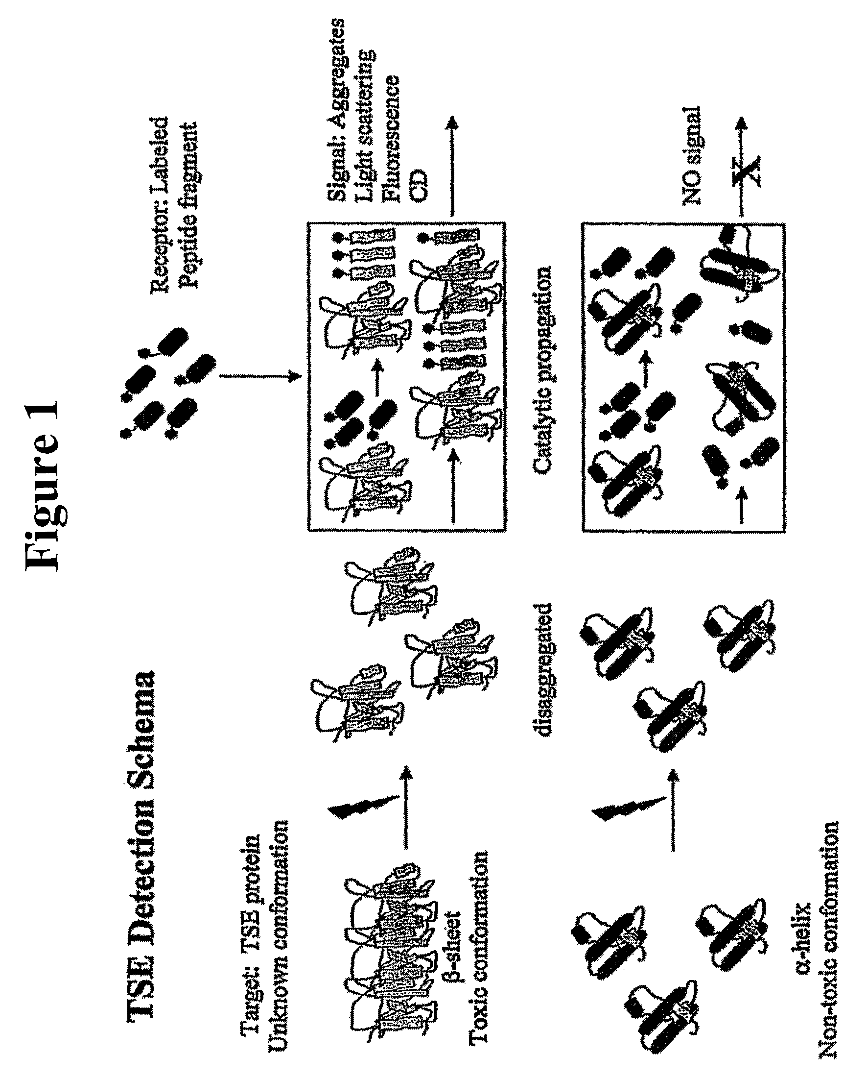
[0091]A conformational propagation assay for prion protein in a murine brain sample was performed essentially as described in Published U.S. Patent Application 2005 / 0026265 A1, and as depicted in FIG. 1. The assay was performed using the murine-specific pallindromic 33-mer probe depicted below (SEQ. ID. NO. 49), labeled at both ends with a pyrene label. Other probes for human / hamster and sheep / bovine prions are presented below as well, and can be used for detection of prions in those species.

[0092]

H2N-VVAGAAAAGAVHKLNTKPKLKHVAGAAAAGAVV-COOH(murine sequence; SEQ. ID. NO. 53)H2N-VVAGAAAAGAMHKMNTKPKMKHMAGAAAAGAVV-COOH(human / hamster sequence; SEQ. ID. NO. 54)H2N-VVAGAAAAGAVHKMNTKPKMKHVAGAAAAGAVV-COOH(sheep / bovine sequence; SEQ. ID. NO. 55)

For improved labeling, the above sequences have been modified to contain an additional lysine (K) residue at the C-terminus (SEQ. ID. NOs. 56-58) to allow addition of a pyrene label.

[0093]

(SEQ. ID. NO. 56)H...

example 2

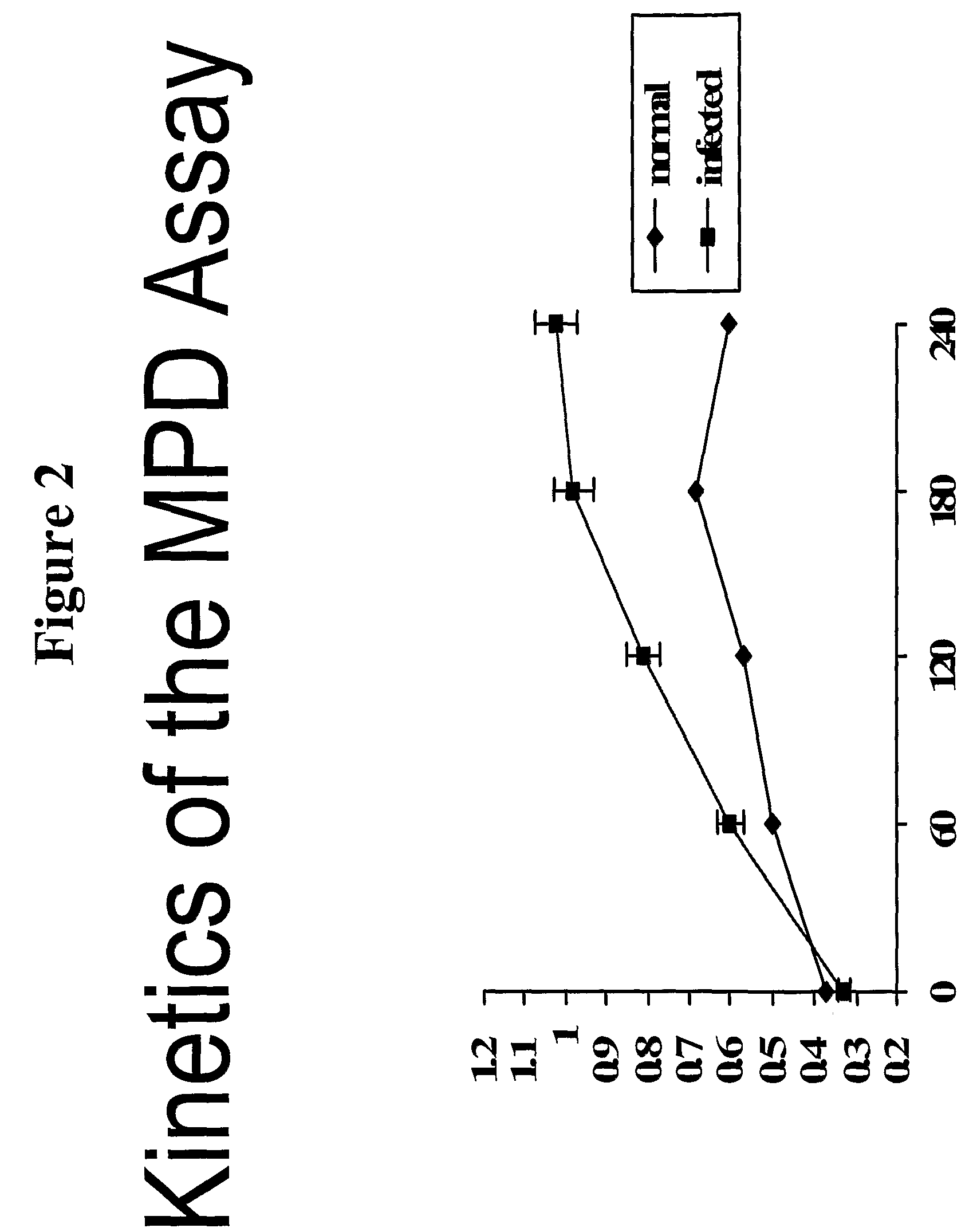
Demonstration of Kinetic Component of the Misfolded Protein Diagnostics (MPD) Assay Using Murine Brain Tissue

[0094]Murine brain tissue from Scrapie-infected and healthy animals was obtained and fractionated on a sucrose gradient. The MPD Assay of Example 1 was performed, using phosphotungstic acid as a precipitating reagent, followed by addition of the murine specific peptide probe. The mixture was allowed to react in Tris:TFE (50:50) mixture per the timecourse shown on the x-axis of FIG. 2. The results show suitable detection of prion protein in samples, which is above background levels for normal brain tissue.

example 3

Fractionation of Prion-Containing Sample According to the Present Invention

[0095]Sheep serum was obtained and mixed with sheep-specific peptide probe (see sequences above), and the mixture was incubated for a sufficient amount of time to permit association of the probe with any prion protein in the serum. The mixture was applied directly to a Sepharose 4B column, and fractions collected. The fractions were analyzed for fluorescence by exciting at 350 nm and scanning from 350-600 nm on a luminescent plate reader. The luminescence of each fraction from healthy and infected sera is depicted in FIG. 3. This data provides for visualization of elution of reactive fractions from the column. As can be seen, using the method of the present invention, not only can the infectious serum be distinguished from the healthy, it can occur in “real-time”. Because the peptide was incubated with the sample prior to fractionation, the assay for misfolded protein is performed during the fractionation.


PUM

PropertyMeasurementUnit
excitation wavelengthaaaaaaaaaa
wavelengthaaaaaaaaaa

Abstract

The invention provides methods and kits for detecting conformationally altered proteins, such as prions or other proteins associated with disease states, in a sample. The methods comprise selectively capturing and separating complexes of peptide and conformationally altered protein from substances that interfere with detection of such complexes, and preferably amplification of the detection signal b addition of a second double-labeled peptide.

Description

CROSS-REFERENCE TO RELATED APPLICATIONS[0001]The present application relies on, and claims the benefit of the filing date of, U.S. provisional patent application No. 60 / 652,733, filed 15 Feb. 2005, the entire disclosure of which is hereby incorporated herein by reference.GOVERNMENT SUPPORT[0002]The inventions disclosed herein were partly funded by grants. Therefore, to the extent that rights to such inventions may accrue to the U.S. Government, the following statement, required under 37 C.F.R. §401.14(f)(4) applies: This invention was made with government support under 5 R44 HL070399-04, awarded by the National Institute of Health. The government has certain rights in the invention.BACKGROUND OF THE INVENTION[0003]The invention relates to the field of detection of proteins in biological samples. More specifically, the invention relates to methods and kits for detecting proteins and prions in a sample containing biological substances, and compositions employed by those methods and ki...

Claims


Application Information

Patent Timeline
Patent Type & Authority Patents(United States)
IPC IPC(8): G01N33/53G01N31/00
CPCG01N33/54333G01N2800/2828
Inventor ORSER, CINDY S.PAN, TAOSETHI, JASMEET
Owner PRESYMPTO INC
Why Eureka
Try Eureka