
Tissue repair devices of rapid therapeutic absorbency

a tissue repair and absorbency technology, applied in the field of soft tissue repair devices, can solve the problems of limited active agent amount that can be added to the implant, ineffective exposure of therapeutic fluid to the side without pores, and difficulty in preloading an implant with active agents, so as to minimize the possibility of tissue adhesion and fast absorb liquid

Active Publication Date: 2013-11-12
ETHICON INC
View PDF23 Cites 128 Cited by
  • Summary
  • Abstract
  • Description
  • Claims
  • Application Information

AI Technical Summary

Benefits of technology

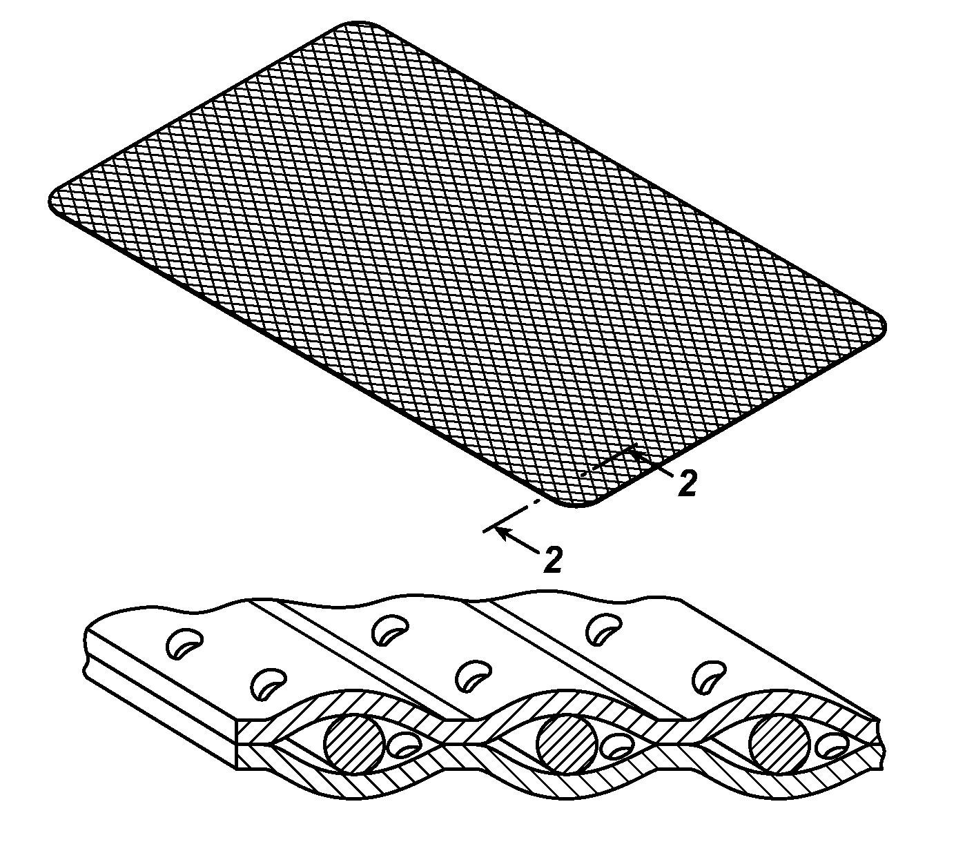
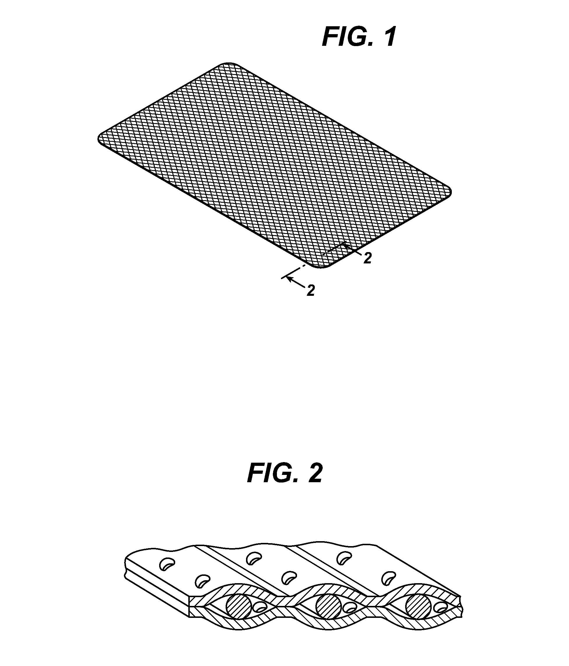
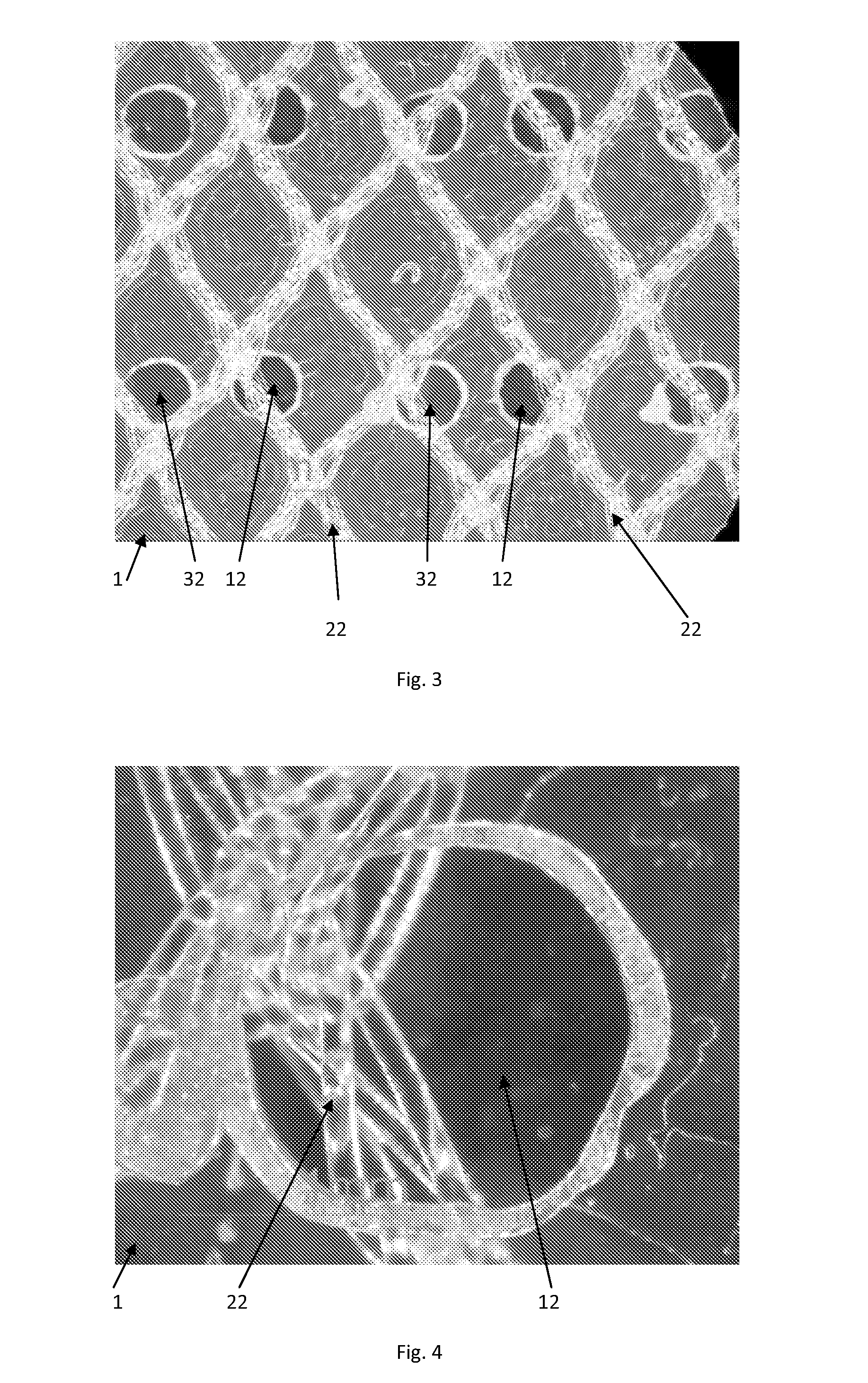
[0010]Accordingly, novel tissue repair implant medical devices are disclosed. The tissue implant medical device of the present invention has a tissue repair member having a plurality of member openings or pores and is preferably a fabric such as a mesh. The repair member has opposed first and second sides. A first polymer film, having first film pores, is mounted on the first side of the member. A second polymer film, having second film pores, is mounted on the second side of the member. The first film pores are not in alignment with the second film pores, that is, the pores are offset, such that tissue-to-tissue contact is substantially prevented.
[0013]The tissue repair devices of the present invention have many advantages. One advantage of the devices of the present invention is to allow an active agent-containing liquid to impregnate the repair fabric and the films in a short period of time, while not exposing facing or adjacent tissue to direct contact once implanted, thereby minimizing the possibility of tissue adhesions. The devices of this invention are particularly well suited to dipping into solutions of active agents, whether in a batch process (such as in an operating room environment) or through a manufacturing process, and demonstrate fast absorption of liquids.

Problems solved by technology

However, preloading an implant with an active agent can be difficult.
In addition, the amount of active agent that can be added to the implant is limited unless the active agent is delivered in a controlled release manner by the implant for controlled release to the adjacent tissues.
If pores are present in only one film layer of the implant, the therapeutic fluid may not be effectively exposed to the side without any pores.
This may result in seroma formation.
Currently marketed and commercially available products that are coated with collagen films (e.g., Parietex®Composite (PCO) MESH) have to be incubated for 5-10 minutes in a solution of active agent, which is relatively time consuming task to perform when in an operating room (OR) setting and while the patient is under anesthesia during a procedure.
A further drawback with current commercially available products is that the active agent coatings are very sensitive to mechanical forces during handling in the operating room, and using forceps to manipulate or place the implants can easily destroy such coatings and may lead to disintegration of the product.
Since both polypropylene and ePTFE both do not accept hydrophilic liquids very well, it is anticipated that the delivery of such meshes along with a coating solution of active ingredient through a trocar to the surgical site would be difficult.

Method used


Image

  • Tissue repair devices of rapid therapeutic absorbency
  • Tissue repair devices of rapid therapeutic absorbency
  • Tissue repair devices of rapid therapeutic absorbency

Examples


example 1

Lightweight Mesh Laminated Between Two Porous Monocryl® Films

[0042]A lightweight polypropylene mesh having the same knitting structure as Ultrapro® brand mesh available from Ethicon, Inc., Somerville, N.J. U.S.A. but without the absorbable Monocryl® filaments-(poliglecaprone 25) was prepared. This mesh was heat laminated between two film layers. The first film consisted of 20 μm thick poliglecaprone 25 Monocryl® film that was extruded and laminated with an 8 μm thick poly-p-dioxanone (PDS) film. The pre-laminate was laser-cut with 1 mm holes or pores with a hole-to-hole distance of 5 mm. A second laminate layer comprising poliglecaprone 25 Monocryl film having a thickness of —————— was laser cut and pre-cut in the same manner as the first laminate layer above. Both films were placed in such a way that the holes or pores were not in alignment (i.e., offset) and were mounted as opposed outer films on the outer surfaces of the polypropylene mesh. The film mesh construct was laminated i...

example 2

Horizontal Dipping

[0048]This example demonstrated the wetting capabilities of the tissue repair devices of the present invention compared with non-porous devices.

[0049]Several 8×12 cm laminates with pore-containing films (pore diameter=1 mm, pore spacing=5 mm) were prepared according to Example 1. Several 8×12 cm laminates with nonporous films were prepared according to Example 1 with the exception that nonporous films were used in place of the porous films. The dry weight was determined for the porous and nonporous laminates.

[0050]The laminates were placed horizontally into a flat vessel containing 500 ml 0.2% Lavasept solution for 10 seconds (made from a Lavasept concentrate (20% PHMB), Lot 7383M03). The laminates were taken out and slightly shaken, to remove excess of liquid and weight was determined again.

Table 1 contains the results of the horizontal dipping experiments.

[0051]

TABLE 1HORIZONTAL DIPPING EXPERIMENTSDryWettedWeightWeights(Before(After 10 sWeightAvgdipping)dipping)i...

example 3

Horizontal Dipping in the OR and Handling Properties

[0055]Laminates were prepared in accordance with Example 1, but in an 18 cm×14 cm size. Pore-containing and nonporous laminates were placed through a conventional 12 mm trocar inserted through to the abdominal cavity of a swine. The implants were easily movable (sliding) at the intestine and then placed against the abdominal wall. Pore-containing and nonporous laminates were removably self-attaching to the abdominal wall. No instrument was needed to keep them up in place.

[0056]The same handling behavior was observed even for 10 second isotonic saline pre-wetted implants.

[0057]With the area weights calculated from Table 1 test articles had an attachment force to the abdominal wall greater than their own area weight of 20 mg / cm2 in the case of the dipped perforated film (calculated from 2 g of the 8×11 cm perforated wet implant in tab 1).

[0058]The devices of this invention were seen to be useful for adhesion prevention as a film barr...


PUM


Abstract

Novel implantable tissue repair medical devices are disclosed. The devices have a central fabric member having anti-adhesion films on both opposed sides. The films have pores, and are arranged such that the pores on the opposed films are offset. The devices are useful in hernia repair procedures.

Description

FIELD OF THE INVENTION[0001]The present invention is directed to soft tissue repair devices capable of minimizing tissue adhesion between adjacent or opposing tissue surfaces, more particularly such soft tissue repair devices capable of rapidly absorbing active agents in an operating room environment prior to implantation.BACKGROUND OF THE INVENTION[0002]Tissue repair or reinforcing implants, such as meshes, may be designed to enable tissue in-growth on one side (e.g., by having open pores or interstices) and resist tissue in-growth on the opposing side (e.g., by having a smooth surface such as a film or non-porous layer, conventionally referred to in this art as an adhesion barrier). This is important when the mesh implants are used or implanted in the abdominal area, for example in hernia repair procedures, where adhesion of the peritoneum (i.e., tissue ingrowth) to the implant is desired while tissue in-growth or adhesions on the visceral side is unwanted (i.e., anti-adhesion). S...

Claims


Application Information

Patent Timeline
Patent Type & Authority Patents(United States)
IPC IPC(8): A61F2/02
CPCA61L27/14A61L27/56A61L31/04A61L31/146A61L31/16A61P31/00A61P41/00A61L31/14A61F2/02
Inventor PRIEWE, JOERG
Owner ETHICON INC
Learn More