Cancer diagnosis marker using the aberrant glycosylation of a protein

a technology of aberrant glycosylation and cancer cells, applied in the detection of post translational modifications, instruments, biochemistry apparatus and processes, etc., can solve the problems of losing information on the glycosylated isoform, experiencing aberrant glycosylation, and losing information on specific structured aberrant glycosylation derived from cancer cells, etc., to facilitate fast and simple diagnosis of cancer

Inactive Publication Date: 2013-11-12
KOREA BASIC SCI INST
View PDF9 Cites 1 Cited by
  • Summary
  • Abstract
  • Description
  • Claims
  • Application Information

AI Technical Summary

Problems solved by technology

However, many glycoproteins or glycolipids located on the surface of cell membrane often experience aberrant glycosylation by the specific signal such as oncogene, etc.
However, this method has a disadvantage of losing information on the glycosylated isoform in relation to characteristics of glycosylation and glycosylated structure in the specific glycosylation site of each protein because this method only enables mass analysis of numbers of glycosylated isoform mixtures composed of all different isoforms originated from different proteins and glycosylation sites but having equal weights.
However, this method has a disadvantage of losing information on specific structured aberrant glycosylation derived from cancer cells because normal glycosylation structure cannot be maintained during glycan-capturing.
If the corresponding antibody is not prepared, the protein cannot be analyzed by this method.
Lectin-blotting utilizes gel-separation technique, suggesting that there is still a disadvantage of limited analyzing speed and questions remain on liability in quantitative analysis.
However, this technique requires a liable antibody and it is very difficult to obtain each antibody against every glycoprotein newly identified at a massive scale.

Method used


Image

  • Cancer diagnosis marker using the aberrant glycosylation of a protein
  • Cancer diagnosis marker using the aberrant glycosylation of a protein
  • Cancer diagnosis marker using the aberrant glycosylation of a protein

Examples


example 1

Sample Preparation

[0087]To overexpress N-acetylglucosaminyltransferase (GnT-V), the glycosyltransferase overexpressed in cancer cells, human colon cancer cells were transfected with MGAT5, resulting in the preparation of stable transfectant cells. Then, the GTN-V overexpressing cells (GnT-V-treated cells) and the control cells were cultured. Equal amounts of culture fluids obtained from both group were concentrated until the volume reached 2 ml respectively. The concentrated samples were reduced using 14 mM β-mercaptoethanol, followed by desalting. One mg of the desalted protein was loaded to L-PHA-avidin-agarose beads, to which phosphate-buffered saline (PBS) was added. The mixture stood at 4° C. for 12 hours. The lectin conjugated protein was washed with PBS three times, and then the protein was separated from lectin by using 6 M urea. The obtained protein was 10-fold diluted with 50 mM ammonium bicarbonate, followed by hydrolysis using 10 ug of trypsin for overnight at 37° C. wit...

example 2

Selection of Marker Candidates Via Peptide Analysis

[0088]HPLC (high-performance liquid chromatography) was performed using trap column (C18, 5 um, 300×5 mm) and analytical column (C18, 5 um, 75 um×10 cm) to analyze the samples prepared in Example 1, followed by LC / ESI-MS / MS using LTQ-FT mass spectrometer (Thermo Finnigan), the electrospray ionization (ESI) mass spectrometer. Each sample protein was hydrolyzed by using trypsin to obtain peptides. Some of the prepared peptide samples were 10-fold diluted, which proceeded to liquid chromatography connected to the mass spectrometer by 10 μl.

[0089]Based on the results of the mass spectrometry, various proteins were qualified from the samples obtained from the GnT-V treated cell line and the normal control cell line by using search engines such as MASCOT and SEQUEST. The relative amount of each protein existed in each sample was calculated based on the frequency of qualitative analysis of each protein. Significant proteins were screened f...

example 3

Confirmation of Marker Glycoprotein by Mass Spectrometry

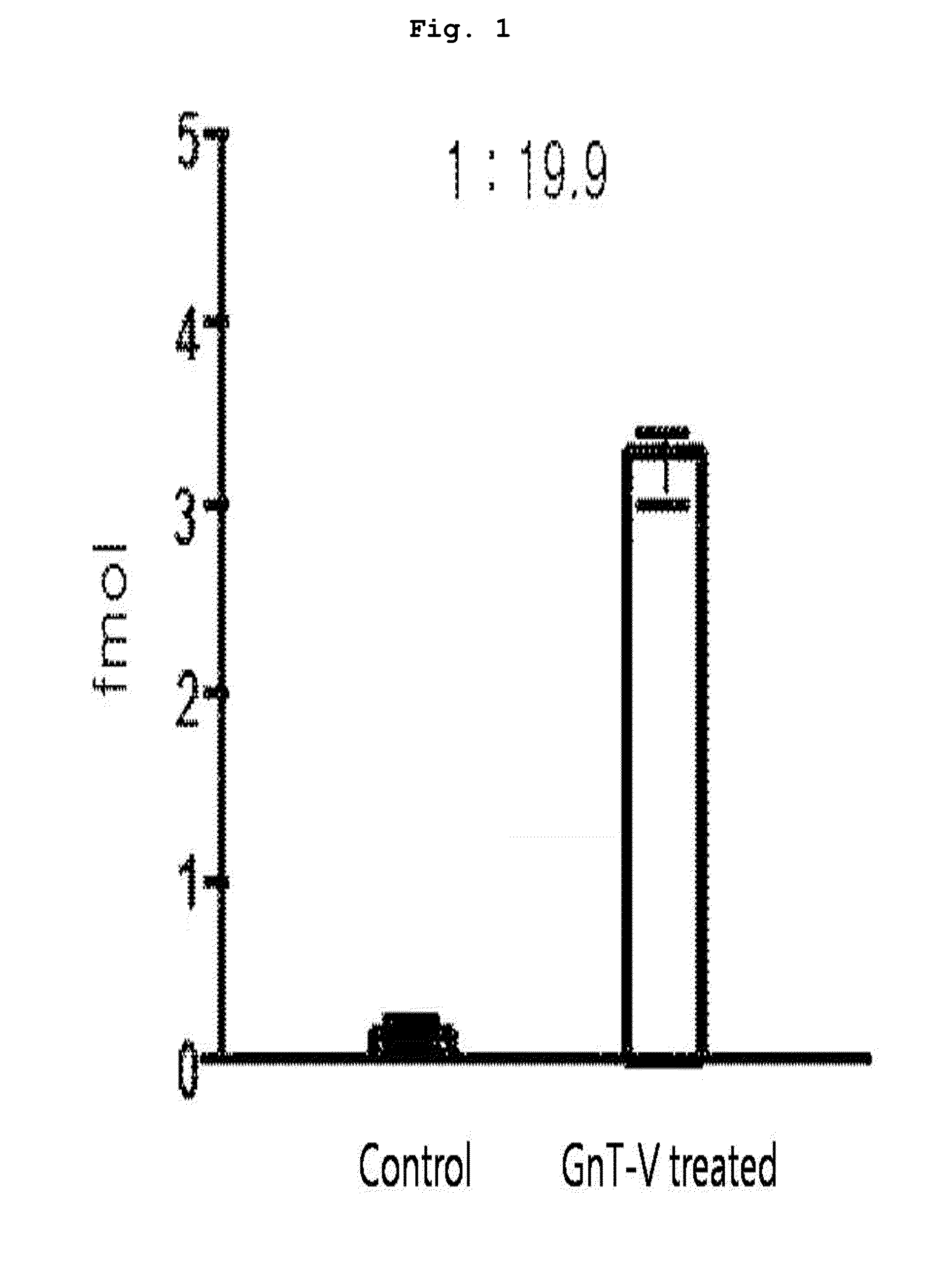
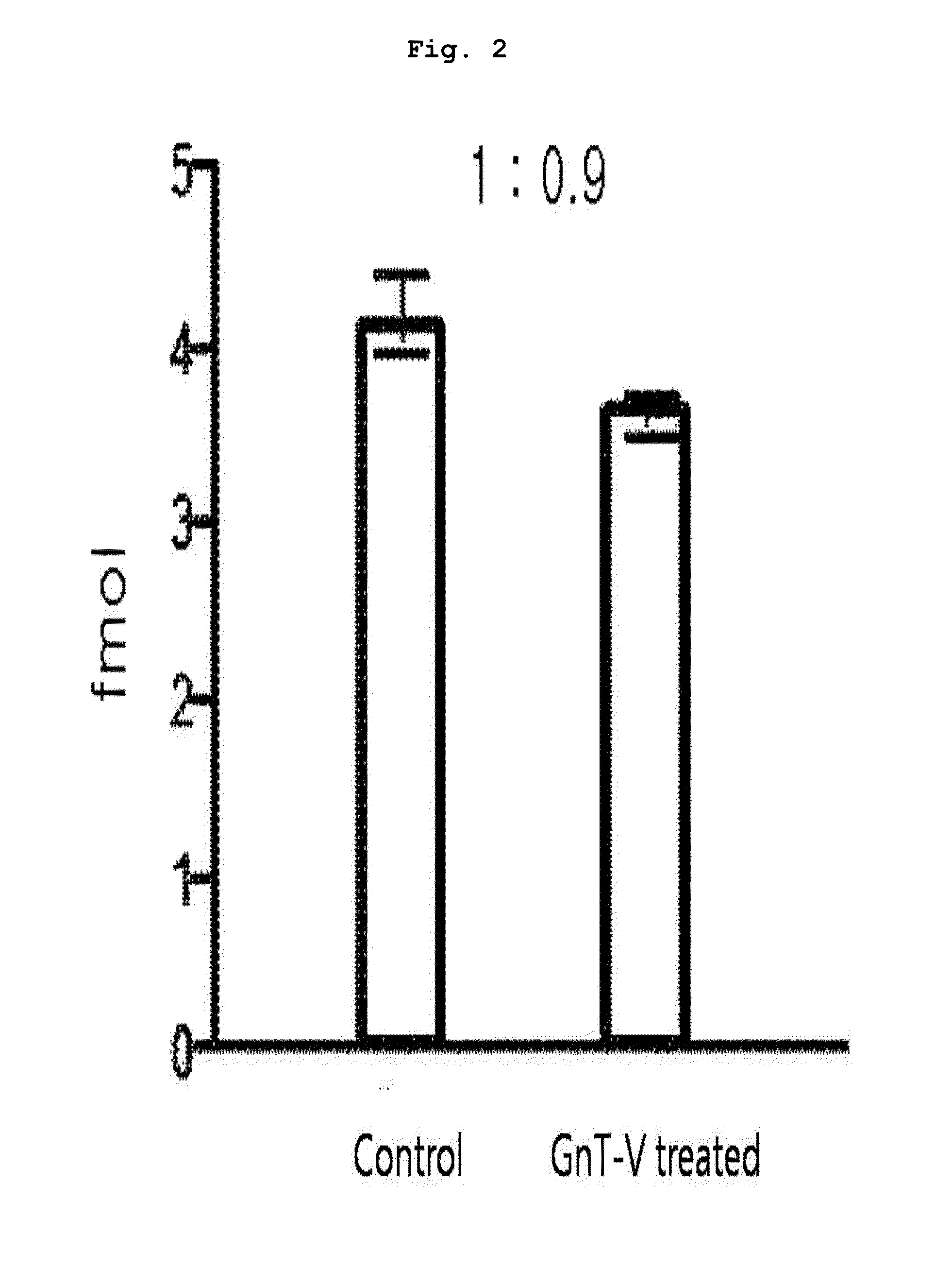
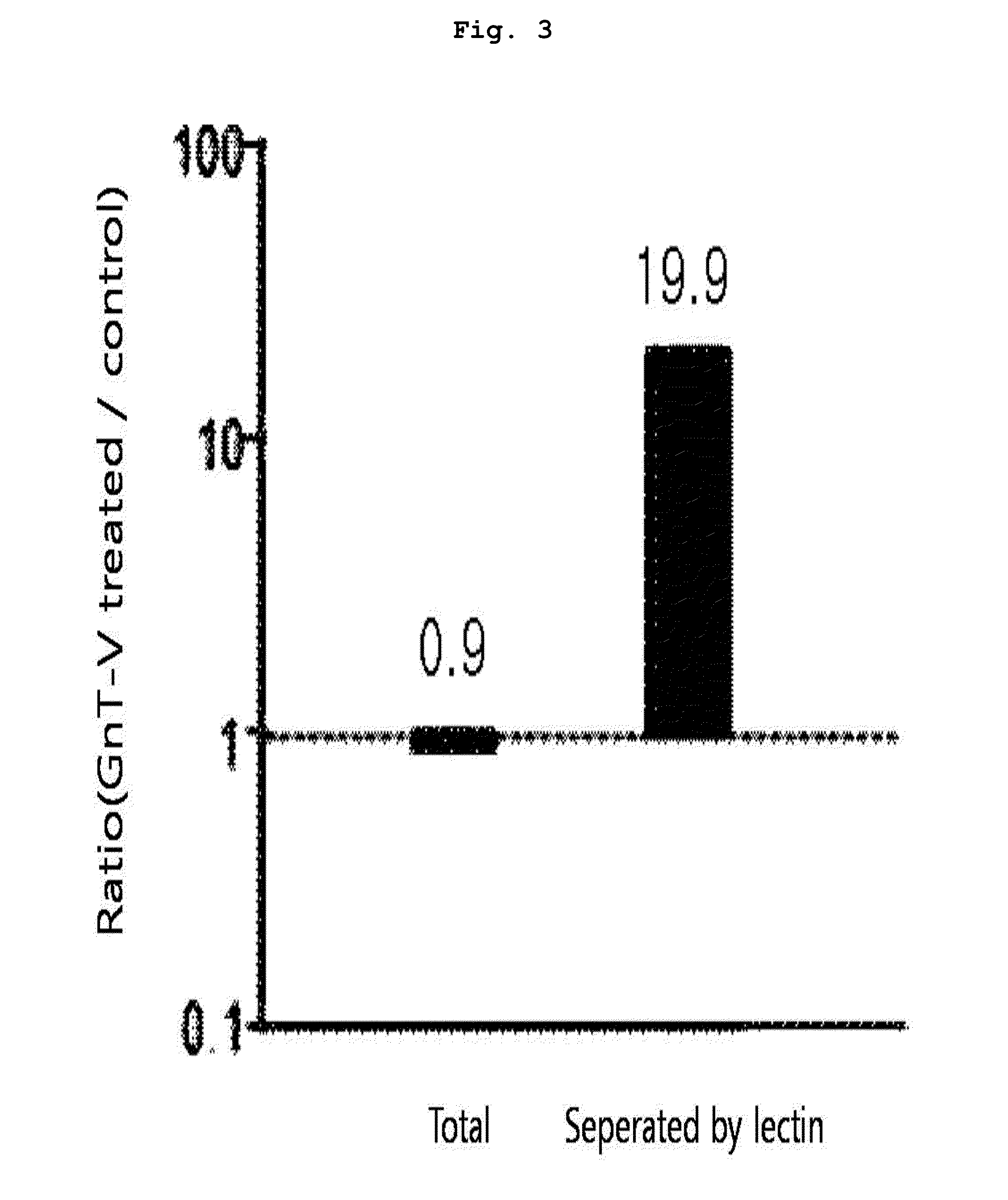
[0093]10 μl of each peptide sample prepared from the protein of each subject in Example 1 was taken, to which 50 fmol of the marker peptide standard material was added to make the total volume 50 μl. 4 samples were prepared; 2 of them were through lectin separation and the other 2 were not finished with lectin separation. The standard material labeled with target peptide isotope was the synthetic standard material that had the identical amino acid sequence with the target peptide (marker peptide) but was different in peptide mass. This material was used as an internal standard material in MRM MS. In quantitative mass spectrometry, calibration curve was made according to the changes of concentrations of the target peptide and the synthetic standard material under the same analysis conditions.

[0094]A part (10 μl) of each prepared sample (50 μl) was used for LC / MRM MS. Quantitative analysis was performed in triplicate with the mar...


PUM

PropertyMeasurementUnit
densityaaaaaaaaaa
volumeaaaaaaaaaa
MWaaaaaaaaaa

Abstract

The present invention relates to a method for diagnosing cancer using information on the aberrant glycosylation of a glycoprotein. More precisely, the present invention relates to a cancer diagnosis peptide marker, which is screened by: a step of separating, using a lectin, a glycoprotein which is aberrantly glycosylated in accordance with the occurrence of cancer; and a step of selecting a hydrolyzed peptide marker derived from the aberrantly glycosylated glycoprotein by observing the quantitative changes in the separated glycoprotein. The present invention also relates to a method for diagnosing cancer using the peptide marker as an agent.

Description

CROSS REFERENCE TO RELATED APPLICATIONS[0001]This application is a national stage application under 35 U.S.C. 371 of PCT Application No. PCT / KR2010 / 009323 having an international filing date of 24 Dec. 2010, which designated the United States, which PCT application claimed the benefit of Korean Application No. 10-2009-0133088 filed 29 Dec. 2009, the entire disclosure of each of which is incorporated herein by reference.BACKGROUND OF THE INVENTION[0002]1. Field of the Invention[0003]The present invention relates to a method for diagnosing cancer by quantitative analysis of a glycoprotein which is aberrantly glycosylated in accordance with the occurrence of cancer.[0004]2. Description of the Related Art[0005]Proteins are involved in various life-supporting activities and post-translationally modified by signal transmission whenever necessary. The most representative post-translational modification processes are glycosylation and phosphorylation. In particular, regarding glycosylation ...

Claims


Application Information

Patent Timeline
Patent Type & Authority Patents(United States)
IPC IPC(8): C12Q1/00G01N33/00G01N33/48G01N33/53
CPCG01N30/7233G01N33/574G01N2030/8831G01N2333/4724G01N2440/38G01N33/68G01N30/72
Inventor AHN, YEONG HEEYOO, JONG SHIN
Owner KOREA BASIC SCI INST
Why Eureka
Try Eureka