Liver cancer methods and compositions

a liver cancer and method technology, applied in the field of liver cancer methods and compositions, can solve the problems of insufficient sensitivity and specificity of existing tumor markers, afp and pivka-ii, which are employed for immunological assays, etc., and achieves simple, accurate and high-throughput results. , the effect of easy automation

Inactive Publication Date: 2015-01-06
ROCHE MOLECULAR SYST INC
View PDF6 Cites 12 Cited by
  • Summary
  • Abstract
  • Description
  • Claims
  • Application Information

AI Technical Summary

Benefits of technology

[0030]The purpose of this invention is to solve problems in screening or monitoring of liver cancer by an immunological assay with conventional tumor markers and an echo test for high risk patients of liver cancer, to discover new tumor markers detectable high-sensitively and high-specifically, even liver cancer in early stage which is difficult to detect by conventional methods, and to provide a simple, accurate and high-throughput method using those markers, and a kit and a reagent thereof. This invention also aims to present a method for liver cancer detection which can easily automate by using blood specimens as measuring targets. Furthermore, utilizing appropriate novel tumor markers, it's also an object of the invention to make a real possibility detection of precancerous lesion and prediction of recurrence after treatment of liver cancer for high risk patients of liver cancer, and detection of malignancy of liver cancer and monitoring of progression of liver cancer with time for liver cancer patients.
[0088]It is possible to detect liver cancer, especially that in early stage, in clinical specimens easily and accurately by the testing method in this invention, and it is expected to help improved outcome from treatment for liver cancer patients by detecting in advance a risk of liver cancer and a risk of recurrence after treatment of liver cancer. As another use, it is expected to provide an indication for decision of treatment course of liver cancer by detecting malignancy of liver cancer and monitoring progression of liver cancer with time.

Problems solved by technology

But the cause is as yet not well known.
However, recurrence rate of liver cancer is extremely high, as virus has been not removed and liver diseases have not been cured even if be treated.
However, the sensitivity and specificity by existing tumor markers, AFP and PIVKA-II, which are employed for immunological assay, are insufficient.
Therefore, it is difficult to detect liver cancer in early stage, most cases have already progressed to advanced cancer when detected, and the 5-year survival relative rate of liver cancer patients is very bad in comparison with other cancers (see nonpatent literature 5).
Meanwhile, recent improvement of instruments in an echo test has enabled us to detect liver cancer in early stage, but it is not suited to screening for high risk patients of liver cancer because of some troubles to take around 30 min for a test, to demand technical skills, to depend on performances of instruments and to remain low penetration rate over general hospitals.
Furthermore, as it is difficult to examine whole liver, especially parts hidden by other organs and inside of liver completely, there is the risk overlook liver cancer.
Thus, there have been no sufficiently satisfactory existing methods for detection of liver cancer, especially that in early stage.
However, the previous results of clinical studies are obtained using tumor tissue DNA after confirmed diagnosis for specimens to analyze, and can not meet a screening use for detection of liver cancer in early stage or risk of liver cancer occurrence.
An assay using tissue is not suited to a routine testing due to complicated manipulation and very low capacity of sample processing.
Furthermore, the sensitivity and specificity are insufficient because of improper selection of genes to analyze and a single gene usage for analysis.
Thus, it has been very difficult to utilize identification of methylation on DNA fragments for cancer diagnosis practically.
Because DNAs from variable tissues or blood cells as well as DNA which is derived from tumor tissue are included in blood, it has been difficult to discriminate DNA derived from a particular tumor tissue by using conventional technologies.

Method used


Image

  • Liver cancer methods and compositions
  • Liver cancer methods and compositions
  • Liver cancer methods and compositions

Examples


example 1

Identification of Liver Cancer Specific Methylation Genes

[0121]Using “Affymetrix Human Genome U95A DNA Chips®”, we selected one hundred and one genes whose expression level specifically and significantly reduced twice and over in tumor tissues, namely whose CpG islands would be most likely to be highly methylated, by comparing gene expression levels between seventy-six cases of tumor tissues and sixteen cases of non-tumor tissues which were excised from liver cancer patients by surgery. Furthermore, twenty-three liver cancer specific methylation gene candidates which actually have CpG islands in their promoter regions were selected from said genes.

[0122]Next, each 1 1 μg of tumor tissue genomic DNA which obtained from twenty cases of liver cancer patients in early stage (liver cancer whose stage classification is stage I or II, and tumor size is below 1.3 to 3.6 cm) was BIS-treated, the regions including CpG islands on the above twenty-three candidate genes using said BIS-treated DN...

example 2

Performance Evaluation of Liver Cancer Specific Methylation Genes with a Small Number of Cases

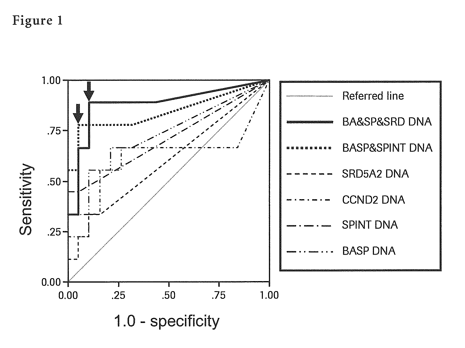
[0126]The total DNA was extracted from 1 ml of serum collected from nine cases of HCV-positive liver cancer in early stage (well-differentiated liver cancer) and nineteen cases of HCV-positive high risk patients of liver cancer (chronic hepatitis and liver cirrhosis) using the DNA Extractor SP Kit for Serum and Plasma (Wako Pure Chemical Industries, Ltd.) and 50 μl of DNA solution was prepared.

[0127]Said DNA solution was BIS-treated using a self-prepared reagent and 50 μl of BIS-treated DNA solution was prepared. Next, each methylation specific PCR (MSP) for seven liver cancer specific methylation genes (BASP1 gene, SPINT2 gene, APC gene, CCND2 gene, CFTR gene, RASSF1 gene and SRD5A2 gene) was performed with 5 μl of said BIS-treated DNA solution. In said MSP, by adding TaqMan probe specific to each gene together, quantification of methylated DNA by real-time PCR was carried out. Quantificat...

example 3

Performance Evaluation of Liver Cancer Specific Methylation Genes with a Large Number of Cases

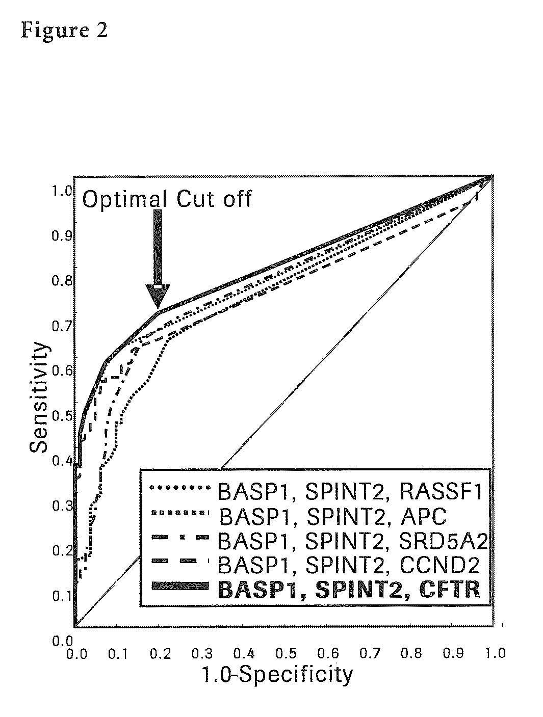
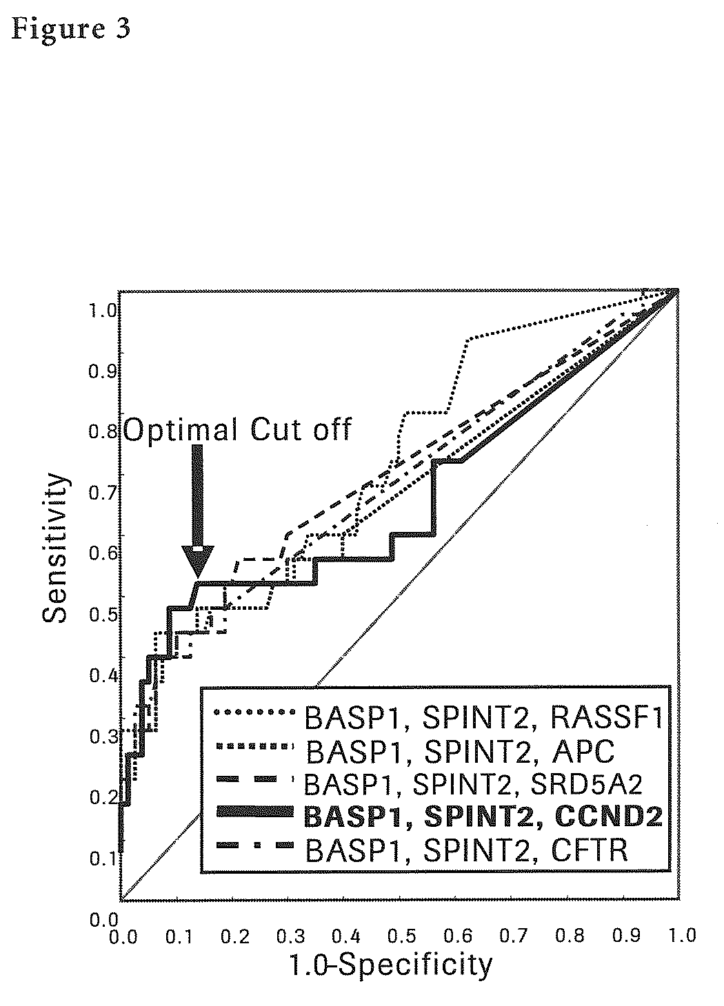
[0139]The total DNA was extracted from 1 ml of serum collected from one hundred and nineteen cases of HCV-positive liver cancer patients (including twenty-five cases of liver cancer in early stage whose stage classification is stage I (stage based on The General

[0140]Rules for the Clinical and Pathologic Study of Primary Liver Cancer, see nonpatent literature 15) and tumor size is below 3 cm, and twenty-one cases of liver cancer in early stage that is well-differentiated) and eighty cases of HCV-positive high risk patients of liver cancer (chronic hepatitis and liver cirrhosis) using the DNA Extractor SP Kit for Serum and Plasma (Wako Pure Chemical Industries, Ltd.) and 50 μl of DNA solution was prepared. Said DNA solution was BIS-treated using a self-prepared reagent and 50 μl of BIS-treated DNA solution was prepared.

[0141]Next, each methylation specific PCR (MSP) for seven liver cancer sp...


PUM

PropertyMeasurementUnit
diameteraaaaaaaaaa
diameteraaaaaaaaaa
sizeaaaaaaaaaa

Abstract

The present invention provides a novel method for detection of liver cancer. This method detects high-sensitively, high-specifically, simply and accurately liver cancer, especially that in early stage by identifying and / or quantifying methylation on particular genes and / or their DNA fragments in clinical specimens, and by combining said methylated DNA values with existing tumor marker values and / or DNA amounts in blood. This invention also detects a precancerous lesion, detects a risk of recurrence after treatment of liver cancer, detects malignancy of liver cancer and monitors progression of liver cancer with time by the same method. As for particular genes, BASP1 gene, SPINT2 gene, APC gene, CCND2 gene, CFTR gene, RASSF1 gene and SRD5A2 gene are mentioned.

Description

[0001]This application is a national stage of the PCT application PCT / EP2008 / 001640, which claims priority to JP Application 2008-021438 filed Jan. 31,2008; and to JP Application 2007-53312 filed Mar. 2,2007.FIELD OF THE INVENTION[0002]The present invention provides a method, a kit and a reagent to detect liver cancer, risk of liver cancer, risk of recurrence of liver cancer and malignancy of liver cancer, and to monitor progression with time of liver cancer by identifying and / or quantifying methylation on particular genes and / or their DNA fragments in clinical specimens.BACKGROUND OF THE INVENTION[0003]Liver cancer develops from chronic liver diseases (chronic hepatitis and liver cirrhosis), and most cases from those with persistent infection of hepatitis virus. In Japan over 95% of liver cancer patients have sustained infection with hepatitis B virus (HBV) and hepatitis C virus (HCV), especially over 80% of those derives from HCV-associates diseases. Generally, patients with hepat...

Claims


Application Information

Patent Timeline
Patent Type & Authority Patents(United States)
IPC IPC(8): C12Q1/68C07H21/02C12P19/34C07H21/04
CPCC12Q1/6886C12Q2600/118C12Q2600/106C12Q2600/112C12Q2600/154C12Q2523/125C12Q2600/156
Inventor HAMAMOTO, YOSHIHIKOIIZUKA, NORIOMIURA, TOSHIAKIMORIBE, TOYOKIOKA, MASAAKITAMATSUKURI, SHIGERU
Owner ROCHE MOLECULAR SYST INC
Why Eureka
Try Eureka