Measurement of autoantibodies at low conductivity with increased sensitivity

a low conductivity, autoantibodies technology, applied in the direction of instruments, biochemistry apparatus and processes, material analysis, etc., can solve the problems of low avidity of dsdna/anti-dsdna antibody complexes, limits the detection of autoantibodies with relatively high avidity, and only as useful as the quality of the data they produce, so as to achieve enhanced sensitivity, less constraints on detection thresholds, and low conductivity

Active Publication Date: 2015-07-07
TAKEDA PHARMA CO LTD
View PDF8 Cites 4 Cited by
  • Summary
  • Abstract
  • Description
  • Claims
  • Application Information

AI Technical Summary

Benefits of technology

[0015]The present invention provides a low conductivity buffer system and methods of using same to achieve autoantibody detection with enhanced sensitivity. The increased sensitivity permits detection of autoantibodies even at concentrations as low as 0.2 μg / mL, thereby placing less constraints on the detection threshold and bypassing various problems attendant with assaying more concentrated samples, i.e., less diluted samples. Moreover, the buffer system and methods of the invention significantly increase sensitivity of the immunodetection assays without a corresponding loss in selectivity. The low conductivity conditions also provide a fluidic environment of low conductivity in which binding to non-antigen coated wells is dramatically reduced.

Problems solved by technology

Results from an autoantibody assay, however, are only as useful as the quality of the data it produces.
Farr assays, for instance, precipitate immune complexes of dsDNA / anti-dsDNA Abs at high salt concentrations in ammonium sulphate, which causes low avidity dsDNA / anti-dsDNA antibody complexes to dissociate and thereby limits detection to autoantibodies with relatively high avidity.
This result is a setback as autoantibodies are highly heterogeneous with respect to their avidity and those of moderate to low avidity may also have clinical significance, as seen in Alzheimer's disease (AD) where IgG autoantibodies bind to epitope on beta amyloid (Aβ) monomers and aggregate with moderate avidity.
Furthermore, Farr assays employ a radiolabel and are both labor-intensive and expensive.
IFA Crithidia, while capable of detecting autoantibodies of moderate to high avidity, is fairly time-consuming and subjective due to its dependence on slide scoring.
ELISA assays are generally more sensitive and susceptible to automation and, thus, often the assay of choice.
They are generally less specific than other methods such as immunopreciptation and immunoelectrophoresis and suffer from a host of issues as documented in the literature.
Despite recent advances in Alzheimer's research, efforts to accurately measure anti-amyloid-β autoantibodies have been undermined by many obstacles.
One obstacle is the existence of multiple classes of human antibodies that recognize linear as well as conformational neo-epitopes on aggregated forms of Aβ.
The measurement of low-avidity, polyreactive autoantibodies is a particular challenge due to high background binding to empty ELISA wells and assay interference from other plasma proteins and components in the blood samples.
Results were less than ideal as the depletion was not specific to autoantibodies which bind blank plates and depletion reduced the already low concentrations of anti-Aβ autoantibodies, which further complicated measurement.
The low-avidity of polyreactive autoantibodies makes them difficult to detect, and the resulting signal intensity is further affected by interference from other plasma proteins and components, leading to underestimates of the anti-amyloid activity.
This technique, however, increases both signal strength and noise.
At the other end of the spectrum, recent methods directed at improving signal strength of the low-avidity, polyreactive autoantibody detection have resulted in over-estimates of anti-amyloid activity.
Reliable assays for low-avidity, polyreactive autoantibodies have largely proven elusive.

Method used


Image

  • Measurement of autoantibodies at low conductivity with increased sensitivity
  • Measurement of autoantibodies at low conductivity with increased sensitivity
  • Measurement of autoantibodies at low conductivity with increased sensitivity

Examples


example 1

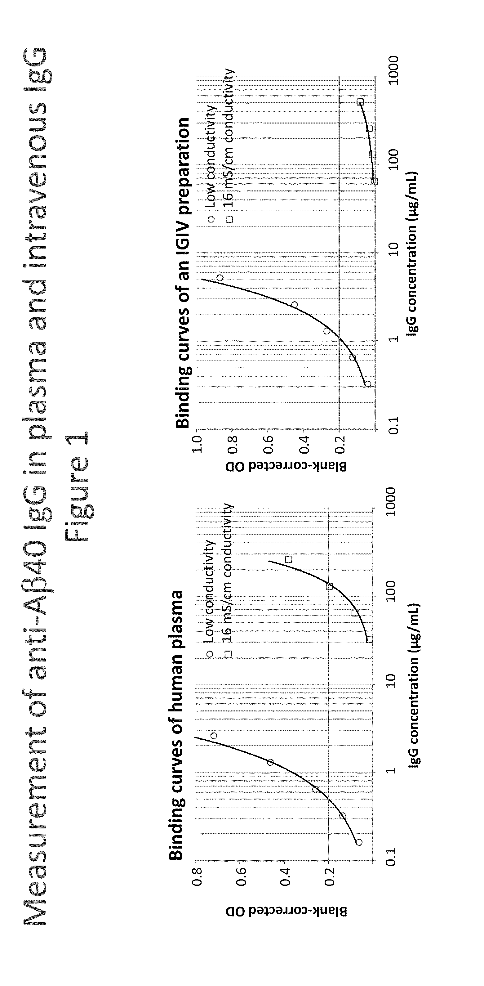
Measurement of Anti-Amyloid(1-40) (Aβ40) IgG in Plasma and in an Intravenous IgG Preparation (IVIG)

[0156]Synthetic human Aβ40 peptide (Calbiochem) was dissolved at 1 mg / mL in trifluoroacetic acid and diluted to 10 μg / mL with 0.1 M NaHCO3—Na2CO3 buffer, pH 9.5. This solution was incubated with the wells of a NUNC Maxisorp F96 plate at 4° C. overnight (100 μL / well). The plate was then washed with washing buffer having 16 mS / cm conductivity or low conductivity concentration of NaCl. In both cases the washing buffer was 20 mM HEPES, 2 mM CaCl2, 0.1% Tween 20 containing 0 (WBs) or 150 mM NaCl (WBi). The samples, a human reference plasma preparation #1R92 and an intravenous IVIG preparation (Gammagard Liquid, #LE12G036), were diluted using the respective washing buffer after addition of 10 mg / mL human serum albumin (hSA). These dilutions buffers were also used for blocking the plate, which was achieved by incubation at room temperature (RT) for 2 h. Then the plate was washed and afterward...

example 2

Measurement of anti Aβ42 IgG in intermediates of IVIG

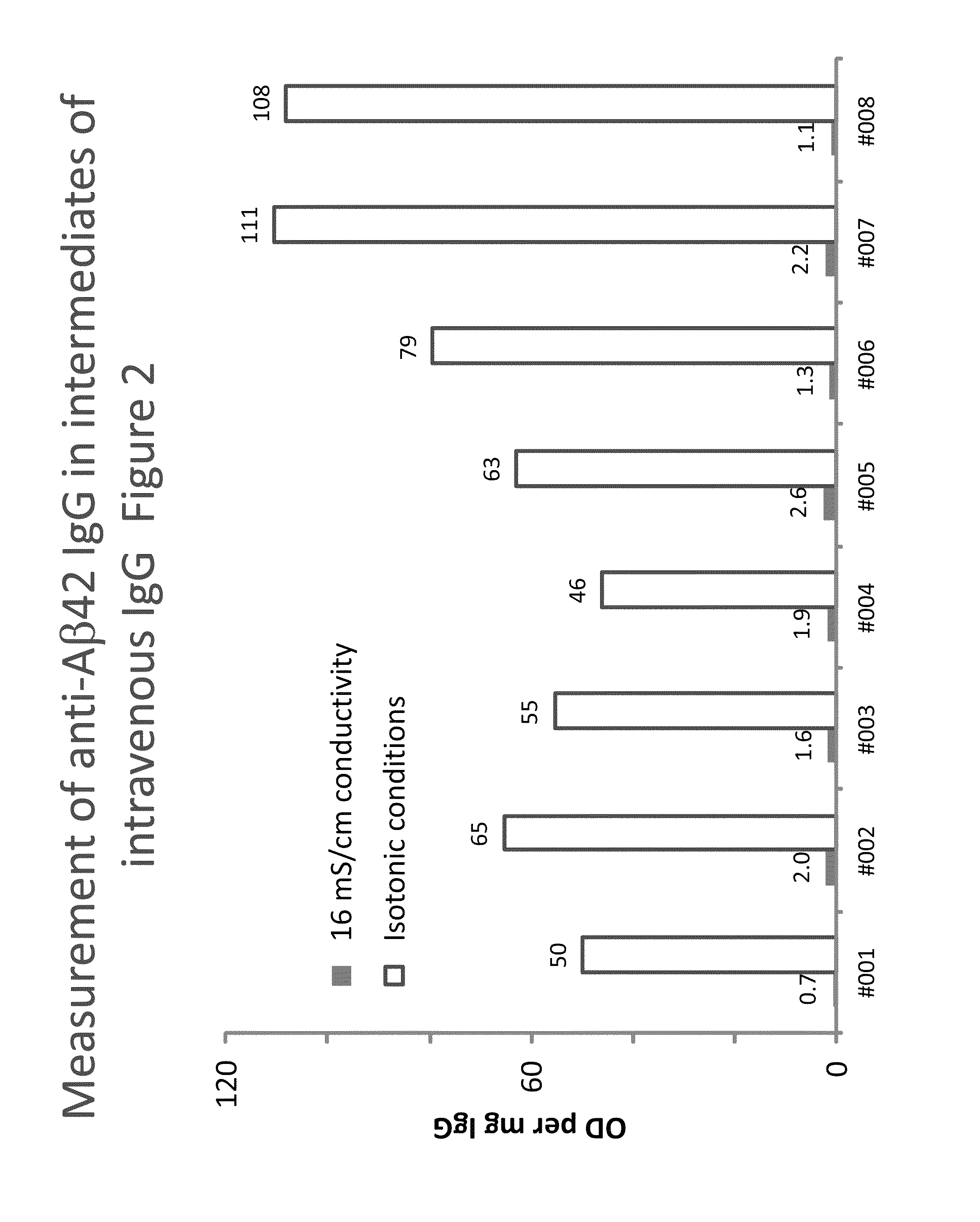
[0158]Eight lots of a process intermediate from the IVIG manufacturing process with a purity of higher than 85% were investigated for their anti-Aβ42 IgG levels using the methods described in Example 1. In contrast to the buffers described the buffers used did not contain CaCl2. FIG. 2 shows the anti-Aβ42 IgG levels measured, expressed as blank-corrected OD per mg of IgG.

[0159]In all cases, the low conductivity conditions resulted in clearly increased signals. The assay signal obtained under low conductivity conditions was on average 50-times higher than that obtained under 16 mS / cm conditions. Individual increases ranged from 24 to 102 times. Concomitantly, the ratio between the signals on Aβ-coated and blank wells decrease by more than 50% on average and we found mean ratios of 0.13 and 0.04 for the 16 mS / cm or low conductivity conditions.

example 3

Measurement of anti Aβ40 oligomer (CAPS) IgG in intermediates of IVIG

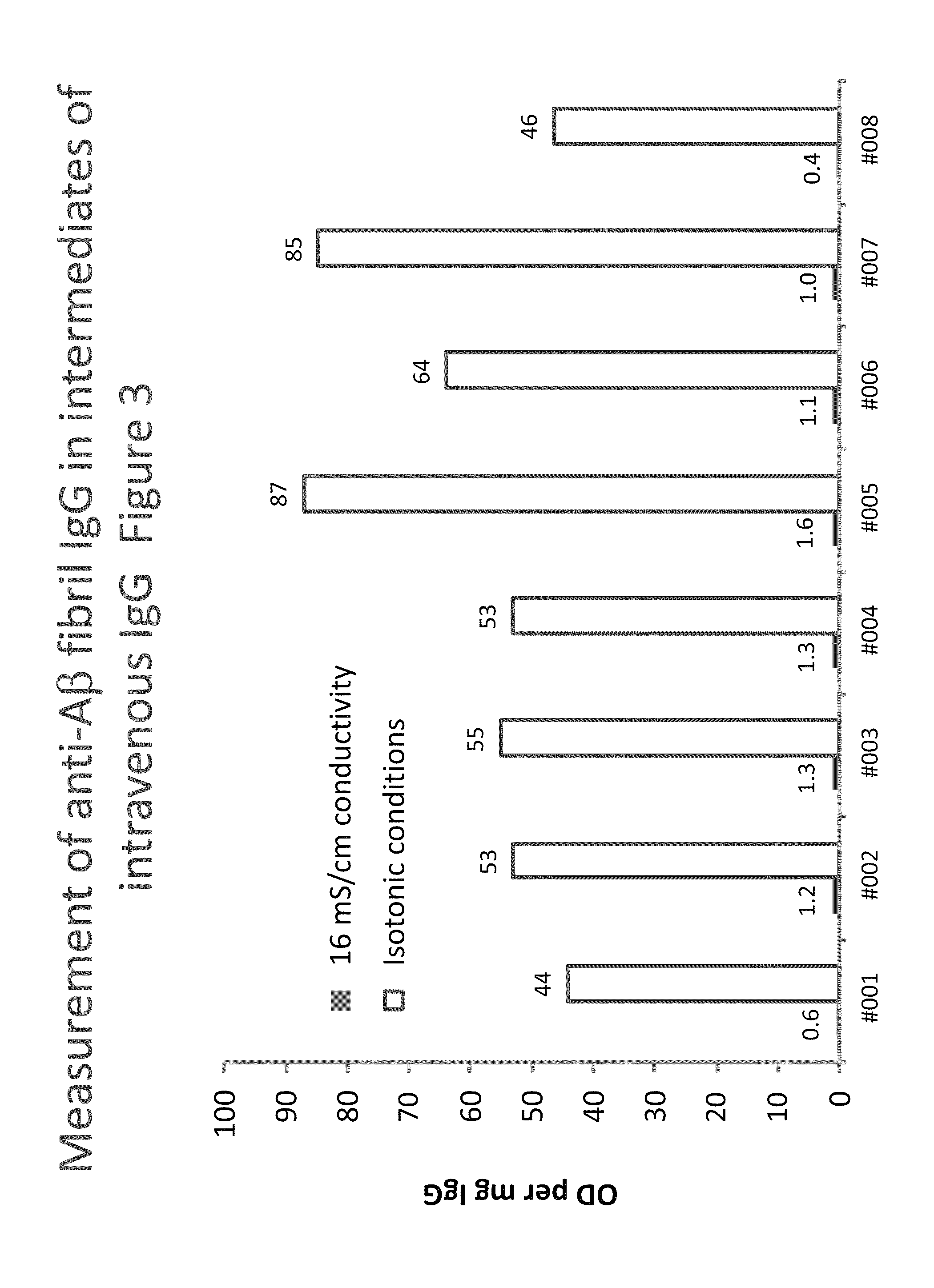
[0160]Plates were coated with anti-Aβ40 oligomers (CAPS, cross-linked β-amyloid protein species), obtained as described (O'Nuallain B. et al., (2010): J Clin Immunol May; 30 Suppl 1:537-42). The assay was then done as described in Example 1 using buffers without CaCl2. Eight lots of a process intermediate from the IVIG manufacturing process with a purity of higher than 85% were investigated for their anti-CAPS IgG levels. Table 2 shows the results giving the IgG concentrations (in μg / mL) and the corresponding signals obtained on the CAPS-coated (OD) and the blank wells (Blank). In addition, the ratio (Ratio) and the difference (Δ) between the corresponding signals and the signal normalized to the protein concentration, expressed as OD / mg are given:

[0161]

TABLE 2Measurement of anti-CAPS IgGSampleIgG (μg / mL)ODBlankRatioΔODOD / mg16 mS / cm conditions#0016541.1070.0750.071.0331.58#0024211.0860.0950.090.9912.36#0035951.4180...


PUM

PropertyMeasurementUnit
concentrationsaaaaaaaaaa
conductivityaaaaaaaaaa
ionic strengthsaaaaaaaaaa

Abstract

Methods for detecting or capturing low-avidity autoantibodies in a biological sample are provided. Target antigen used to assay for the low-avidity autoantibodies of interest is immobilized on a solid phase. The biological sample is contacted under low conductivity condition with the target antigen for which the autoantibodies has specific binding affinity. Binding of the target antigen to the autoantibodies of interest in the biological sample is then detected to ascertain the presence or concentration of the autoantibodies of interest.

Description

[0001]The present application claims the benefit of U.S. Provisional Application No. 61 / 570,211, filed Dec. 13, 2011, which is expressly incorporated herein by reference in its entirety for all purposes.BACKGROUND OF THE INVENTION[0002]The role of autoantibodies in diseases is a subject of intense research. Autoantibodies form a diverse repertoire with the capacity to target antibody, peptidic, or nucleic acid self-antigens. These autoantibodies may be monovalent or polyreactive and may exhibit varying affinities and avidities; dissociation constants can range from 10−5 to 10−8 M. While autoantibodies are widely implicated in autoimmune diseases, a subset of autoantibodies also plays a non-pathological role in maintaining immunological homeostasis and a properly functioning immune system. There is mounting evidence that autoantibodies are essential to the survival and / or development of T and B lymphocytes in the peripheral immune system, defense against microbial infections, inflamm...

Claims


Application Information

Patent Timeline
Patent Type & Authority Patents(United States)
IPC IPC(8): G01N33/564G01N33/68G01N33/543
CPCG01N33/6854G01N33/54393G01N33/564G01N2333/4709
Inventor WEBER, ALFREDENGELMAIER, ANDREASCHWARZ, HANS-PETER
Owner TAKEDA PHARMA CO LTD
Why Eureka
Try Eureka