Method and system of ultrasound scatterer characterization

a scatterer and ultrasound technology, applied in the field of methods and systems of ultrasound scatterer characterization, can solve the problems of unsuitable methods for all applications, unsuitable for characterizing dense concentrations of scatterers, and risk of strok

Active Publication Date: 2016-07-12
VAL CHUM PARTNERSHIP
View PDF11 Cites 10 Cited by
  • Summary
  • Abstract
  • Description
  • Claims
  • Application Information

AI Technical Summary

Benefits of technology

[0010]An object of the present invention is to provide an improved method and system for ultrasound scatterer characterization. The present invention reduces the difficulties and disadvantages of the aforesaid prior art by providing a novel approach to characterizing scatterers based on ultrasound scattering or backscattering measurements which may be automated and operated in real-time.
[0018]In another embodiment, modelling the ultrasound data includes consideration of an attenuation of the scatterers and the medium, the method further comprising estimating the attenuation at the same time as estimating the physical property value of the scatterers. Simultaneously estimating the attenuation and the physical property value can comprise minimizing a cost function which is a mean quadratic difference between the ultrasound data provided and the modelled data. In this embodiment, the ultrasound data as a function of the spatial organization parameter is based on the Born approximation and the spatial organization parameter is a structure factor. The ultrasound data may represent an ultrasound backscatter coefficient (BSC). In this case, the physical parameter value to be estimated can be the packing factor W and the diameter D of the scatterers, the method comprising applying:
[0026]Advantageously, the method is not limited to the quantification of ultrasound data e.g. backscatter coefficient (BSC) and its frequency dependence (spectral slope, y-intercept, mid-band fit), unlike known prior art methods which can only provide ultrasound related parameters that are difficult to interpret and are also intrinsically dependent on the frequency that is used. The present approach, on the other hand, allows these ultrasound related parameters to be transformed into physically representative quantitative data, e.g. absolute values of a packing factor W and an average cluster (aggregate) non-dimensional size D. Embodiments of the present invention therefore allow comparisons of studies in different frequency ranges and comparison with results from other fields of study.
[0029]For example, an embodiment of the present invention can be used to monitor red blood cell aggregation which can be a marker of inflammation. Inflammation may be encountered in patients with obesity, diabetes, atherosclerosis, rheumatoid arthritis, rheumatic fever, bacterial infection, meningitis, neonatal illness and pneumonia, inter-current sepsis, septic shock, cancer, heart disease, those at risk of developing deep vein thrombosis and pulmonary embolisms (e.g., hospitalized immobile patients), and those undergoing cancer treatment and surgery, such as heart surgery, and patients with HIV. The inflammatory response accompanied by abnormal levels of red blood cell aggregation can lead to an acute vaso-occlusive crisis or thrombotic complications which may be fatal. Platelet aggregation occurring in response to a blood vessel injury can also be monitored with ultrasound and characterized by an embodiment of the present invention. Monitoring and measuring red blood cell aggregation may also be valuable in assessing the response to therapy and changes in the pathological state of many chronic inflammatory diseases that are difficult to monitor clinically. This is particularly true of rheumatoid arthritis, polymyalgia, giant cell arthritis, inflammatory bowel disease, systemic vasculitis, chronic migraine, antibacterial therapy, genetic blood diseases as beta-thalassemia and sickle cell anaemia, malaria, and future coronary events in patients with unstable angina or after coronary occlusion or post-surgery. Aggregation of red blood cells and their disaggregation (unclustering) can therefore be detected and monitored in vivo as red blood cell clusters are moving in a blood vessel. In this way, bedside monitoring of red blood cell aggregation in patients in an emergency or in intensive care units to allow immediate diagnosis and treatment is possible. Self-monitoring is also possible.

Problems solved by technology

However, these methods are not suitable for all applications.
They are particularly unsuitable for characterizing dense concentrations of scatterers in a medium such as dense suspensions of particles.
Also, the presence and size of embolisms in the blood vessels of a patient can be indicative of their risk of suffering a stroke.
However, these known ultrasound techniques cannot provide quantitative or absolute physical parameters or accurate qualitative estimates of the physical parameters of the scatterers in dense medium.
Attenuation can be particularly problematic in the characterization of biological material because of the frequency-dependent attenuation due to intervening tissue layers that distorts the frequency dependence of scattering from the tissue microstructure.
However, this method can result in inaccurate compensation, especially in the case of characterization of biological material as intervening tissue layer attenuations can vary between layers and can also vary between patients.
However, the attenuations of each intervening layer between the ultrasound probe and the site of the scatterers being characterized must be estimated by a separate measurement technique which means that the compensation cannot be in real-time or performed by a single device.
Furthermore, these methods assume strong hypotheses on spatial tissue homogeneity which can lead to inaccuracies.
A further limitation of known prior art ultrasound scatterer characterization techniques is that some of them rely on sampling a bulk suspension to be tested (i.e. “in vitro” methods).
However, these in vitro methods cannot provide real-time analysis as samples from a bulk suspension must be isolated to be analyzed.
This is undesirable in the case of manufacturing processes where the process must be interrupted to take a sample, or where the substance concerned is toxic, caustic, hot or pressurized.
Sampling can also introduce contamination into the bulk or can dilute the bulk.
Also, it can introduce sampling errors if the sample taken is not indicative of the bulk.
In the case of medical applications, such as detecting red blood cells or other particulates in blood samples, there is the obvious drawback of having to take a blood sample from the patient and the associated health and safety issues for both the patient and the person taking the blood.

Method used


Image

  • Method and system of ultrasound scatterer characterization
  • Method and system of ultrasound scatterer characterization
  • Method and system of ultrasound scatterer characterization

Examples


example 1

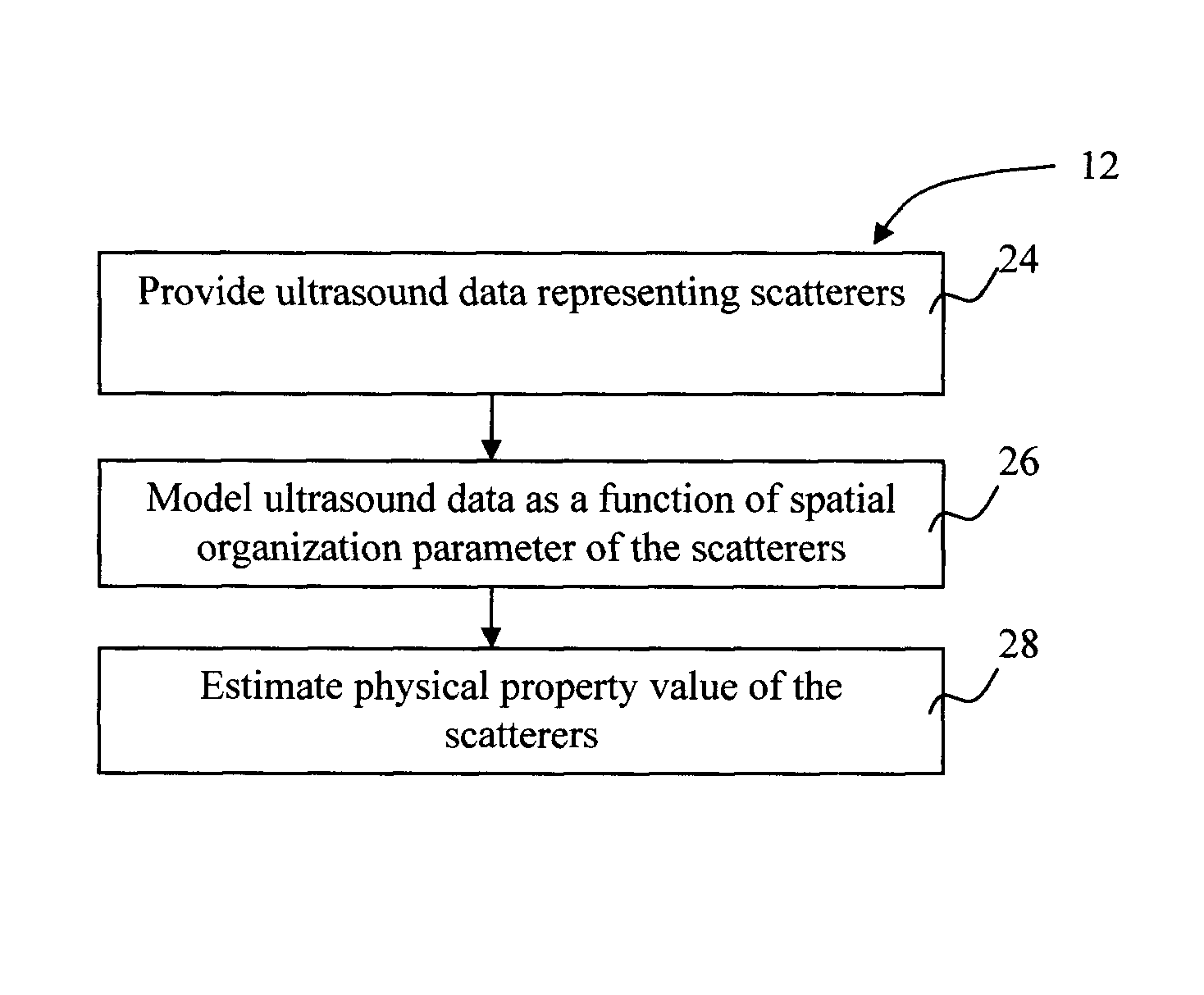
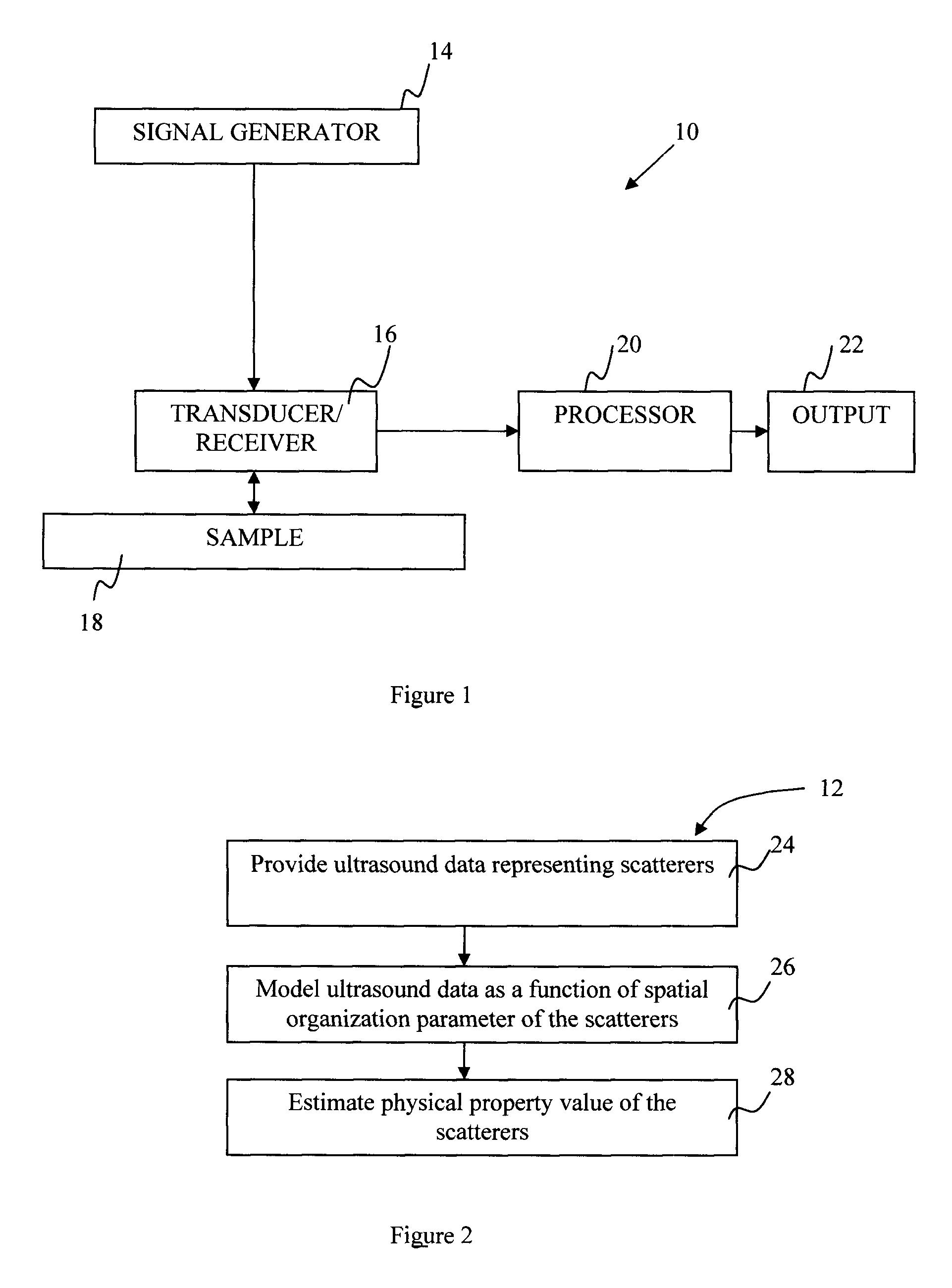
This Example Illustrates a Validation of the First Embodiment of the Method 12

Summary:

[0129]Different samples of whole blood and red blood cell suspensions at 6% and 40% hematocrits were prepared and quantitatively characterized at room temperature using three focused wideband transducers covering the bandwidth from 9 to 30 MHz. According to the first embodiment of the present invention, a second order Taylor approximation of the structure factor was used to achieve data reduction of the BSC measurements, to extract two physical parameters, the packing factor (W) and the mean normalized dimension of isotropic aggregates (D). The D parameter was validated by an optical imaging method at 6% hematocrit under static conditions. Both parameters closely matched theoretical values for non-aggregated red blood cells. This therefore validated the theoretical SFSE model of the present invention.

Blood Preparation:

[0130]Fresh porcine whole blood was anti-coagulated with 3 g / L of ethylene diamin...

example 2

This Example Illustrates In Vivo Results Obtained by the First Embodiment of the Method 12 to Obtain Information about Red Blood Cell Aggregation in a Brachial Vein of a Normal Subject

[0155]A high frequency transducer (Visualsonics, Canada) was used to image the brachial vein of the normal subject's forearm. In this case, the RMV-710 probe was used (25 MHz center frequency, focal length 1.5 cm). The probe was positioned parallel to the vein of interest to provide a longitudinal section view of the vein. Each image contained 384 vertical lines. The equivalent to 10 images of the same section were digitized at a sampling frequency of 500 MHz. A region of interest was delimited around the venous valve. The data was processed as described for the first embodiment of the method 12, except that the power spectra were calculated on windows of 32 points positioned at 10 points intervals, for an overlap of 22 points. This allowed a 2D mapping of W and D over the image. Each estimate of W and...

example 3

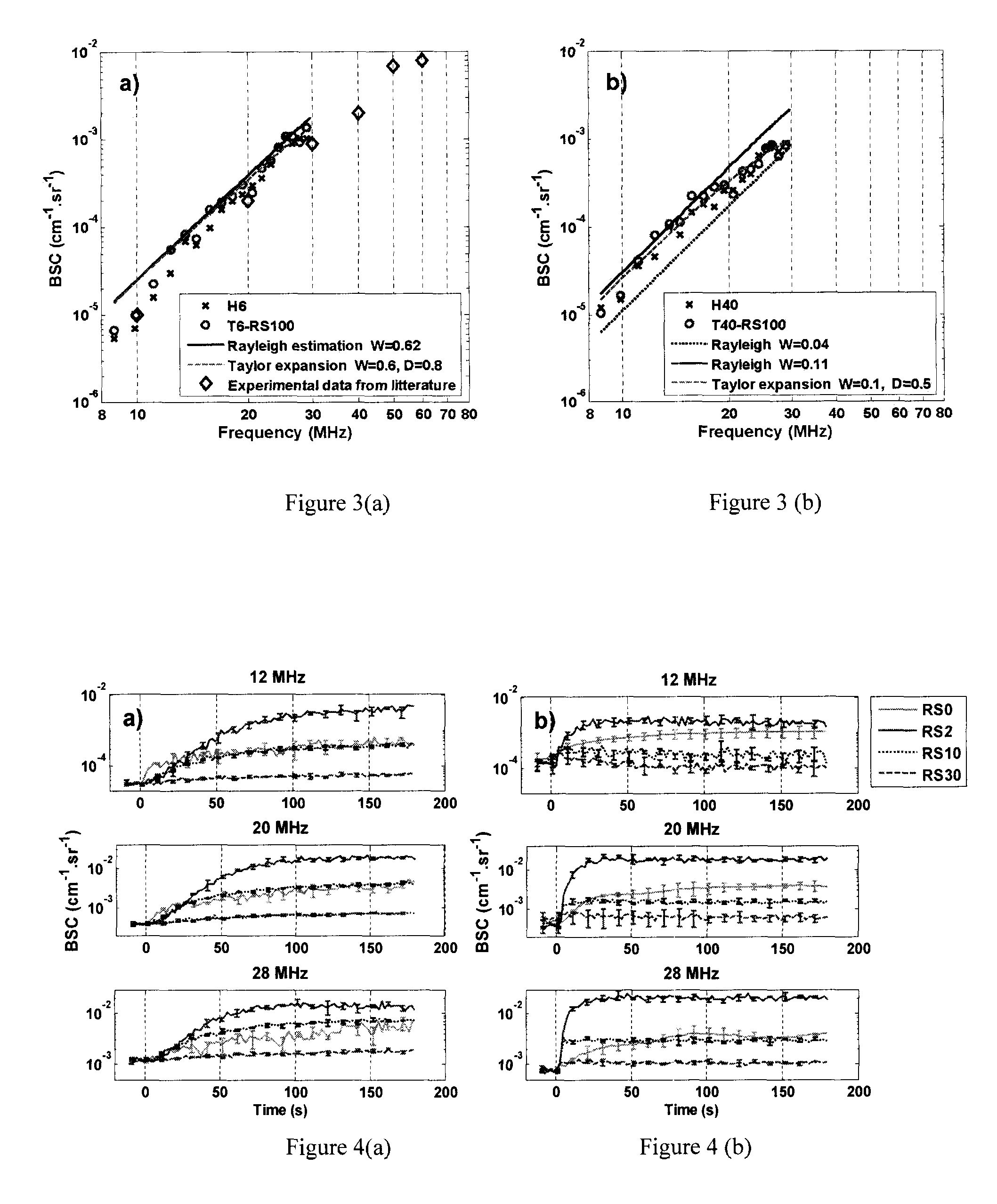
This Example Illustrates In Vitro Results Obtained from the Second Embodiment of the Method 12 where Attenuation and the Physical Parameters are Estimated Simultaneously with the SFSAE Model

Blood Preparation:

[0157]Fresh porcine whole blood was obtained from a local slaughter house, centrifuged and the plasma and buffy coat were removed. Two blood samples were then prepared: (i) a H6 reference sample, which was a 6% hematocrit non-aggregating red blood cells resuspended in physiological saline solution; and (ii) a 40% hematocrit T40 test sample, which consisted of red blood cells resuspended in plasma to promote aggregation.

In Vitro Experiment in a Couette Flow System (Couette Device):

[0158]Ultrasound measurements were first performed in a Couette device to produce a linear blood velocity gradient at a given shear rate (FIG. 17). The system consists of a rotating inner cylinder with a diameter of 160 mm surrounded by a fixed concentric cylinder of diameter 164 mm. A 60 mL blood sampl...


PUM

PropertyMeasurementUnit
frequenciesaaaaaaaaaa
volumeaaaaaaaaaa
frequencyaaaaaaaaaa

Abstract

A method for characterizing ultrasound scatterers in a medium comprising providing ultrasound data representing a region of interest comprising a plurality of scatterers in a medium, the plurality of scatterers including clusters of scatterer sub-units, the scatterers having a physical property value to be estimated and the scatterer sub-units having at least one known physical parameter value; modelling the ultrasound data using an at least second order function of a spatial organization parameter defining the spatial organization of the scatterers; and estimating the physical property value of the scatterers from the modelled ultrasound data and the at least one known physical parameter of the sub-units by a regression of the spatial organization parameter as a function of frequency. A system for characterizing ultrasound scatterers is also included.

Description

CROSS-REFERENCE TO RELATED APPLICATION(S)[0001]The present application is a National Stage Entry of PCT / CA2008 / 000673 filed on Apr. 10, 2008 which claims priority on U.S. Provisional Patent Application No. 61 / 014,991, filed on Dec. 19, 2007, U.S. Provisional Patent Application No. 60 / 991,431, filed on Nov. 30, 2007, and U.S. Provisional Patent Application No. 60 / 911,738, filed on Apr. 13, 2007, all incorporated herein by reference.FIELD OF THE INVENTION[0002]The present invention generally relates to a method and a system of ultrasound scatterer characterization.BACKGROUND OF THE INVENTION[0003]Ultrasound techniques are commonly used as non-invasive or non-destructive diagnostic tools in a range of industries including medicine, foodstuffs, pharmaceuticals, petrochemicals, chemicals and materials processing. In known ultrasound methods, ultrasound echoes are transmitted to scatterers in a medium and backscattered or scattered echoes are detected. Ultrasound parameters such as backsc...

Claims


Application Information

Patent Timeline
Patent Type & Authority Patents(United States)
IPC IPC(8): A61B8/00G01N29/06A61B8/06A61B8/13G01N29/44G01S7/52
CPCG01N29/069A61B8/06A61B8/13G01N29/4472G01S7/52036G01N2291/014G01N2291/02408G01N2291/02836G01N29/4418G01N2291/02416G01N2291/02466
Inventor CLOUTIER, GUYYU, FRANCOISFRANCESCHINI, EMILIESAVERY, DAVID
Owner VAL CHUM PARTNERSHIP
Why Eureka
Try Eureka