
Anterior segment drug delivery

a technology of anterior segment and drug delivery, which is applied in the field of anterior segment drug delivery, can solve the problems of blurring the patient's vision for a prolonged time period, the drug's presence in the bloodstream may have harmful side effects on the rest of the body, and the drug's most wasteful us

Active Publication Date: 2016-08-23
FORSIGHT VISION5 INC
View PDF256 Cites 16 Cited by
  • Summary
  • Abstract
  • Description
  • Claims
  • Application Information

AI Technical Summary

Benefits of technology

The patent is about a medical device that is used to treat glaucoma (a condition that causes high pressure in the eye). The device has two structures: a first structure that acts as a skeletal frame and maintains the position of the implant in the eye for at least 30 days, and a second, cushioning structure that helps to prevent the implant from moving around. The first structure remains a constant size and shape, and it is designed to fit snugly around the inferior and superior conjunctival fornices of the eye. This helps to keep the device in place and maintains its effectiveness in treating glaucoma.

Problems solved by technology

Eye drops and gels can be effective drug delivery vehicles, but can also have significant disadvantages.
The absorption into the bloodstream can have at least two adverse effects: first, most of the drug is wasted and, second, the presence of the drug in the bloodstream may have harmful side effects on the rest of the body.
Gels may adhere more effectively to the eye, but can also blur the patient's vision for prolonged time periods.
Factors that may have contributed to the limited acceptance of prior ocular inserts include their lack of comfort, their propensity for displacement or movement around the eye, their excessive incidents of inadvertent expulsion during sleep or rubbing of eyes, their interference with vision, and / or the difficulty in placing and removing the known drug delivery implants.

Method used


Image

  • Anterior segment drug delivery
  • Anterior segment drug delivery
  • Anterior segment drug delivery

Examples


example 1

Calculation of a Drug's Therapeutically Effective Dosage for an Ocular Insert—Olopatadine

[0107]The drug Olopatadine, for treatment of allergic conditions, will demonstrate a method that can be used in calculating a drug's therapeutically effective plant-delivered dosage based on a drop-administered treatment regimen for that drug. The calculation method involves the following steps: 1.) determining the number of drops desired per application; 2.) multiplying the number of drops by 30 uL (the volume of one drop); 3.) determining the amount of solid drug per uL; 4.) multiplying the results from step 2 by the result from step 3, to find out the amount of solid drug to be applied to the eye on a daily basis; 5.) multiplying the result in step 4 by the number of days of therapy desired for the particular drug; and 6) multiplying by the efficiency of drug delivery. This final amount will preferably be dispersed from an ocular insert.

[0108]Olopatadine is an ophthalmic antihistamine (H 1 -r...

example 2

Drug Delivery Procedure & Elution Rate Control for a Hydrophilic DrugOlopatadine hydrochloride

[0110]Olopatadine HCl (OH) for ophthalmic applications can be formulated as a 0.2% (2 mg / mL) solution. A single 50 uL drop containing 100 ug of OH may be instilled in the eye once a day for 2 weeks. Estimating 5% availability, this gives a 5 ug / day dose delivered to the cornea, for a total of 70 ug over the course of the 2 week treatment. At least 70 ug in dry form could be loaded into the implant and released into the tear film by partitioning the drug reservoir with a membrane (e.g. HEMA, PVA, PVP, GMA, dialysis tubing of cellulose, etc) or embedding the drug within the implant. The release rate could be controlled by altering the surface area exposed to the tear film to tailor the desired 5 ug / day (0.21 ug / hour), by altering a drug release controlling membrane, or the like. It is again assumed in this calculation that 100% of the targeted dose gets to its target location without being w...

example 3

Drug Delivery Procedure & Elution Rate Control for a Hydrophobic DrugPrednisolone Acetate

[0112]Generally, a 1% Prednisolone acetate suspension (10 mg / mL) is given 2 drops (total of approximately 100 uL volume) 4 times daily for a week. Working with the estimate that 5% of a dose is actually available for absorption into the cornea, this amounts to 20 ug / day of Prednisolone acetate. A week's available dose is then 140 ug. The solubility of Prednisolone acetate in aqueous solutions is approximately 240 ug / mL. At least 140 ug of solid Prednisolone acetate could be loaded into the implant, allowing the Prednisolone acetate to dissolve into the tear layer at a rate of about 0.83 ug / hour. The rate could be controlled by the porosity of the implant as well as the surface area exposed to the tear film.

[0113]For these simplified calculations, it has been assumed that 100% of the dose hits the target (the cornea) and is absorbed completely and not lost by tear layer flow away from the cornea...


PUM

PropertyMeasurementUnit
diameteraaaaaaaaaa
arc angleaaaaaaaaaa
diameteraaaaaaaaaa

Abstract

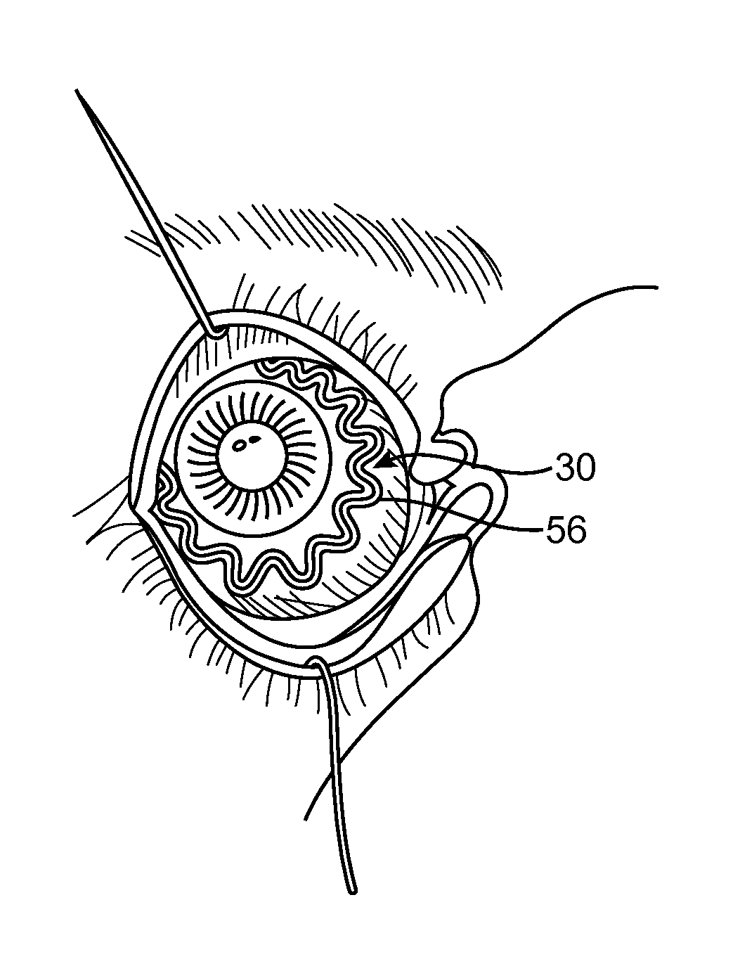
A therapeutic system comprises an ocular insert placed on a region outside an optical zone of an eye. The ocular insert comprises two structures: a first skeletal structure and a second cushioning structure. The first structure functions as a skeletal frame which maintains positioning of the implant along the anterior portion of the eye and provides support to the second, cushioning structure. This first structure maintains the attachment of the therapeutic system to the anterior portion of the eye for at least thirty days. In some embodiments the first structure remains a constant size and shape, e.g. a ring shape, a ring with haptics, or a curvilinear ring that is confined to and restrainingly engages the inferior and superior conjunctival fornices so as to retain the implant within the tear fluid and / or against the tissues of the eye.

Description

CROSS-REFERENCES TO RELATED APPLICATIONS[0001]The present application is a Continuation of PCT / US2010 / 037268 filed Jun. 3, 2010, which application claims the benefit of U.S. Provisional Application No. 61 / 183,839 filed Jun. 3, 2009; the full disclosures, each of which are incorporated herein by reference in their entirety.BACKGROUND OF THE INVENTION[0002]1. Field of the Invention[0003]The invention relates to structures, systems, and methods for treatment of an eye. Exemplary embodiments provide ocular inserts used for drug delivery, along with methods for using ocular inserts positioned on or near the anterior segment of the eye. The exemplary inserts may be worn along the front surface of the eye outside the optical zone, and can deliver one or more drugs at a safe, therapeutically-effective level for at least thirty days.[0004]2. Background of the Invention[0005]A variety of ophthalmic and non-ophthalmic conditions necessitate administration of various drugs to the eye. Eye drops...

Claims


Application Information

Patent Timeline
Patent Type & Authority Patents(United States)
IPC IPC(8): A61F9/00A61K31/56A61K31/5575A61K31/557A61K31/55A61K31/19A61K31/5415A61K31/5377A61K31/4178A61K31/407A61K31/568A61K31/573A61K31/58A61K31/7036A61K38/00A61M31/00A61K31/43A61K9/00A61M35/00
CPCA61F9/0017A61K31/568A61K31/573A61K31/7036A61K31/4178A61F9/0026A61K31/557A61M31/002A61K31/19A61K31/43A61K31/5415A61K38/00A61K31/58A61K31/407A61K31/5575A61K31/5377A61K31/56A61K9/0051A61K31/55A61K31/335A61P27/02A61F2230/0006A61F2/16A61K9/00A61F2/00
Inventor ALSTER, YAIRDE JUAN, JR., EUGENEREICH, CARY J.BOYD, STEPHENSIERRAALEJANDRO, JOSE D.MACFARLANE, K. ANGELASUTTON, DOUGLAS
Owner FORSIGHT VISION5 INC
Learn More