Ultrasound technology to control the spatial organization of cells and proteins in engineered tissues

a technology of engineered tissues and ultrasound, applied in the field of ultrasound technology to control the spatial organization of cells and proteins in engineered tissues, can solve problems such as the development of an ultrasound standing wave field (uswf)

Active Publication Date: 2017-06-27
UNIVERSITY OF ROCHESTER
View PDF11 Cites 2 Cited by
  • Summary
  • Abstract
  • Description
  • Claims
  • Application Information

AI Technical Summary

Benefits of technology

[0014]Methods of promoting angiogenesis throughout three dimensional engineered tissue are needed to fabricate large vascularized tissues and organs for the field of tissue engineering. Angiogenic cell behaviors are modulated by the spatial arrangement of endothelial cells, the organization of their surrounding extracellular matrix (ECM), and by mechanical force application. The present invention demonstrates that ultrasound technology is capable of controlling all three of these regulatory factors and can induce spatial organization of both cells and proteins and subsequently vascularization within three dimensional engineered tissue constructs. The present invention will allow for the successful reconstitution of viable tissues and organs in vitro, making it possible to repair or replace diseased or injured tissues and organs.

Problems solved by technology

When an ultrasonic pressure wave is incident on an acoustic reflector, the reflected wave interferes with the incident wave resulting in the development of an ultrasound standing wave field (USWF).

Method used


Image

  • Ultrasound technology to control the spatial organization of cells and proteins in engineered tissues
  • Ultrasound technology to control the spatial organization of cells and proteins in engineered tissues
  • Ultrasound technology to control the spatial organization of cells and proteins in engineered tissues

Examples


example 1

Experimental Set-Up for Ultrasound Exposures

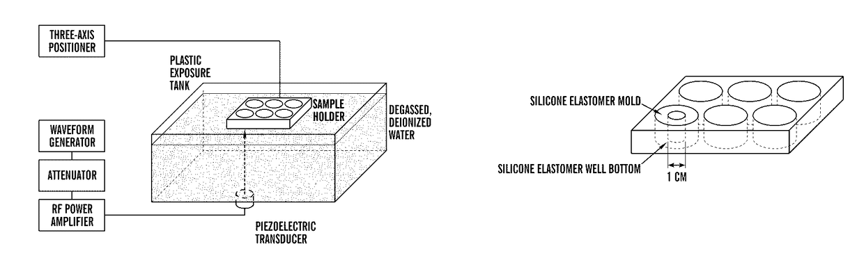
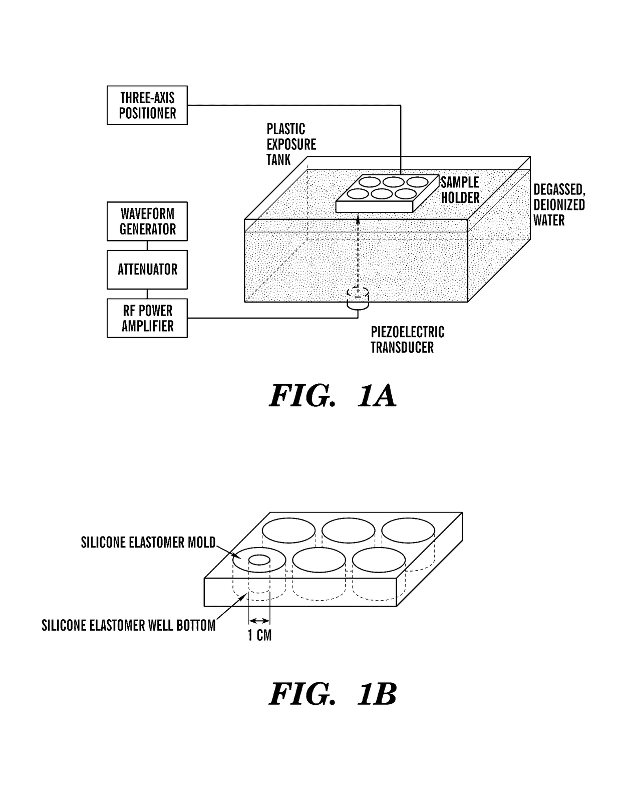
[0088]To investigate the effects of ultrasonic mechanical forces on the promotion of angiogenesis, an ultrasound (US) exposure system was developed in which cells and 3D tissue constructs are subjected to an ultrasound traveling wave field (UTWF) or an ultrasound standing wave field (USWF) (FIG. 1A). Samples are contained within the wells of a modified silicone elastomer-bottomed cell culture plate (FIG. 1B). UTWF are created by minimizing reflections at the sample / air interface with a rubber absorber. USWF are produced by removing the rubber absorber such that the air interface promotes the interference of incident and reflected waves (Blackstock D T, FUNDAMENTALS OF PHYSICAL ACOUSTICS, (Wiley & Sons, 2000), which is hereby incorporated by reference in its entirety). No significant differences in UTWF or USWF are found in the presence of the sample holder compared to without the holder, indicating that the experimental set-up will not int...

example 2

Characterization of Ultrasound Fields

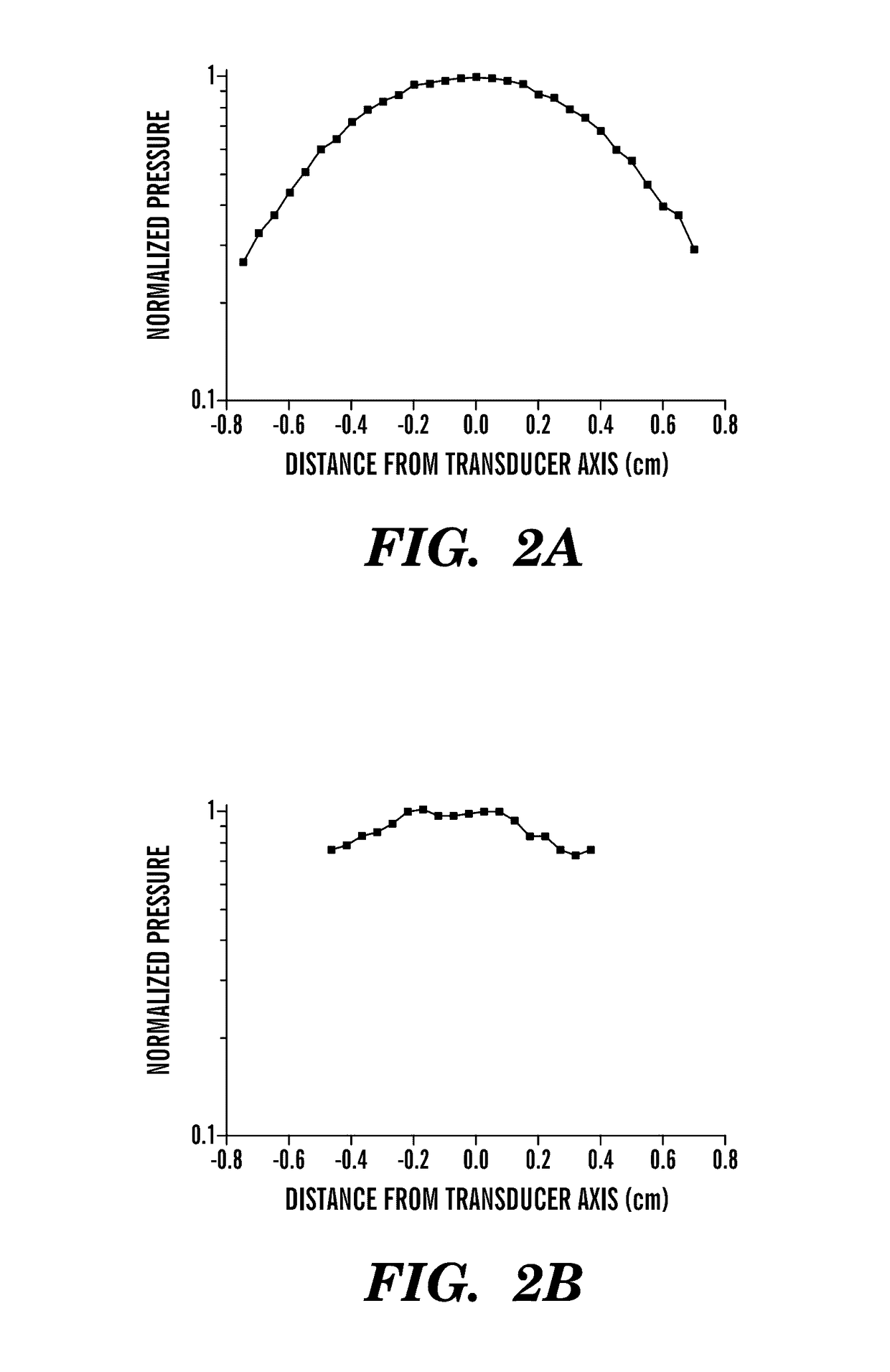
[0091]USWF and traveling wave fields were measured in a water tank and within the sample space using the set-up described above. Resulting spatial distributions of pressure are illustrated below in FIGS. 2A-2D. An unfocused, 1 MHz, 1 inch diameter, piezoelectric transducer was used to produce the US field and a needle hydrophone was used to measure the acoustic pressure.

[0092]For traveling wave fields, transaxial spatial distributions in pressure were measured in the far field, 12.2 cm from the transducer. In the absence of the sample holder, −3 dB and −6 dB beam widths were 0.8 cm and 1.2 cm, respectively (FIG. 2A). The beam pattern within the sample space was not significantly altered from the free field pattern indicating that the sample holder does not interfere with sound propagation (FIG. 2B). The −3 dB beam width remained 0.8 cm, but the −6 dB beam width could not be calculated, indicating that samples will be exposed to a relatively unifo...

example 3

USWF Control of the Spatial Arrangement of Cells Within a 3D Tissue Construct

[0094]The organization of endothelial cells into multicellular assemblies affects angiogenic endothelial cell behaviors (Korff et al., “Integration of Endothelial Cells in Multicellular Spheroids Prevents Apoptosis and Induces Differentiation,”The Journal of Cell Biology 143(5):1341-1352 (1998) and Ino et al., “Application of Magnetic Force-Based Cell Patterning for Controlling Cell-Cell Interactions in Angiogenesis,”Biotechnology and Bioengineering 102(3):882-890 (2009), which are hereby incorporated by reference in their entirety). Exposure of cell suspensions to USWF can result in cellular aggregation at areas of minimum acoustic pressure (the pressure nodes) (Dyson et al., “The Production of Blood Cell Stasis and Endothelial Damage in the Blood Vessels of Chick Embryos Treated with Ultrasound in a Stationary Wave Field,”Ultrasound in Medicine and Biology 1:133-148 (1974), which is hereby incorporated by...


PUM

PropertyMeasurementUnit
frequencyaaaaaaaaaa
acoustic frequencyaaaaaaaaaa
acoustic pressure amplitudeaaaaaaaaaa

Abstract

The present invention is directed to methods of inducing spatial organization of cells an in vitro culture system using ultrasound technology. The invention is further directed to methods of inducing extracellular matrix remodeling and neovessel formation in an in vitro culture system and generating vascularized engineered tissue constructs using ultrasound technology.

Description

[0001]This application claims the priority benefit of U.S. Provisional Patent Application Ser. No. 61 / 179,646, filed May 19, 2009, which is hereby incorporated by reference in its entirety.[0002]This invention was made with government support under grant number R01EB008368 awarded by the National Institutes of Health. The government has certain rights in this invention.FIELD OF THE INVENTION[0003]The present invention is directed to methods of inducing cellular spatial organization, extracellular remodeling, and neovessel formation in an in vitro tissue culture system using ultrasound technology.BACKGROUND OF THE INVENTION[0004]Tissue and organ transplantation is a worldwide therapeutic approach for end-stage organ failure (Nasseri et al., “Tissue Engineering: An Evolving 21st-Century Science to Provide Biological Replacement for Reconstruction and Transplantation,”Surgery 130:781-784 (2001)). Currently, over 100,000 people are in need of an organ transplant in the United States alo...

Claims


Application Information

Patent Timeline
Patent Type & Authority Patents(United States)
IPC IPC(8): C12N13/00C12N5/077C12N5/00C12N5/071
CPCC12N5/069C12N5/0062C12N5/0656C12N2521/10C12N2533/52C12N2533/54C12M35/04A61K9/00A61K35/12C12N13/00C12Q1/02C12Q1/68
Inventor DALECKI, DIANEHOCKING, DENISEGARVIN, KELLEY
Owner UNIVERSITY OF ROCHESTER
Why Eureka
Try Eureka