Keratoprosthesis, and system and method of corneal repair using same

a corneal and keratoprosthesis technology, applied in the field of corneal repair, can solve the problems of scarring of the cornea and cornea, severe damage to the cornea, limited management, etc., and achieve the effect of improving soft tissue adhesion and less extrusion

Active Publication Date: 2018-05-22
UNIV OF MIAMI
View PDF182 Cites 1 Cited by
  • Summary
  • Abstract
  • Description
  • Claims
  • Application Information

AI Technical Summary

Benefits of technology

[0011]The present invention is directed to a novel keratoprosthesis and a system and method for implementing the same in repairing severe corneal damage. The keratoprosthesis comprises a support made of a biocompatible material, preferably titanium, and is disposable fully anterior of the cornea of an eye within a pocket of non-ocular tissue placed thereon. Moreover, the present invention enables improved soft tissue adhesion, which results in less extrusion, than previously known keratoprostheses for treating severely damaged cornea. It also obviates many of the complications and drawbacks of known keratoprostheses. For instance, use of the present keratoprosthesis presents no oral defects.
[0012]More in particular, the keratoprosthesis of the present invention comprises a support, also termed a skirt, which may be made entirely of titanium or of a laminate or composite having outer surfaces of titanium. An optic member is disposed through the support in vision facilitating relation to the eye. The support also includes a plurality of apertures located along the periphery of the support to facilitate securing of the keratoprosthesis in position upon implantation. The support may also comprise a plurality of openings disposed throughout the support, which may be contoured, to allow for tissue integration as well as nutrients and hydration to permeate the keratoprosthesis for overall eye health and prolonged successful implantation.
[0013]A collar extends from the support and surrounds the optic member such that any surface of the keratoprosthesis that comes into contact with the surrounding environment is titanium. These titanium surfaces are pre-treated to create a microstructure that promotes the adhesion of the surrounding soft tissue thereto which forms a bioseal with the keratoprosthesis, preventing epithelial encroachment and resulting extrusion. For example, the titanium surface may be pre-treated with sandblasting and / or acid etching to produce texture on the surface to which the soft tissue may attach and adhere. A locking member may also be disposed between the optic member and collar to secure the optic member to the titanium support.

Problems solved by technology

One of the greatest challenges in ophthalmology is the management of the severely damaged cornea.
These conditions can lead to scarring of the cornea and corneal blindness that is not amenable to treatment or repair by common techniques such as corneal transplantation (also called “penetrating keratoplasty”).
In these circumstances, management is limited to a keratoprosthesis.
The Alphacor™ keratoprosthesis was abandoned in the United States due to poor results after two years, mainly caused by retraction of the cornea at the interface with the prosthesis which can cause aqueous leaks and consequent loss of intraocular pressure and potential endophthalmitis.
The Boston Keratoprosthesis® has had better results but also suffers from cornea thinning and retraction at the cornea-implant interface which, if not repaired in time, can also lead to extrusion, or rejection of the implant, and endophthalmitis.
Human tissue availability might be problematic in countries where donor tissue is difficult to obtain or not available.
In addition, as the Boston Keratoprosthesis® has a fixation member, a plastic or a metal ring, located in the anterior chamber of the patient, contact with the iris is problematic as when the patient's iris closes in bright light, it may contact the fixation ring or the protruding posterior surface of the implant and iris inflammation occurs.
If the membrane is not removed surgically, the increase in intraocular pressure may cause glaucoma and loss of vision.
The major problem with all alloplastic keratoprostheses is the lack of tissue attachment, and therefore a potential rejection.
Despite its relative success, the MOOKP suffers from multiple complications and drawbacks.
In fact, a primary cause of keratoprosthesis failure is extrusion, in which epithelial cells infiltrate the implant area and literally push the implant out.
Secondary infections resulting from bacterial invasion upon epithelial infiltration is also a major problem contributing to prosthesis rejection.
Dental complications arising from the resection of the osteo-odonto lamina may produce damage to adjacent teeth, oro-nasal and oro-antral communication, infection, sinusitis, and nerve damage.
Drawbacks of the MOOKP procedure include a poor cosmetic outcome of the eye.
Although a shell can be developed to cover the buccal mucosa in an attempt to improve the cosmesis of the eye, often times the osteo-odonto lamina or skirt displaces such a large volume of space that the eye cannot be appropriately covered.
Therefore, the appearance of the eye cannot be fully or completely improved.
In another drawback, the MOOKP procedure creates a surgical defect of the oral cavity from the removal of tooth and bone.
However, such an oral defect is complex and generally is difficult to reconstruct.
Finally, the multistage nature of the MOOKP procedure, involving two different groups of surgeons, and the duration of the various steps makes it an inherently prolonged procedure with numerous opportunities for complications to arise.

Method used


Image

  • Keratoprosthesis, and system and method of corneal repair using same
  • Keratoprosthesis, and system and method of corneal repair using same
  • Keratoprosthesis, and system and method of corneal repair using same

Examples


Embodiment Construction

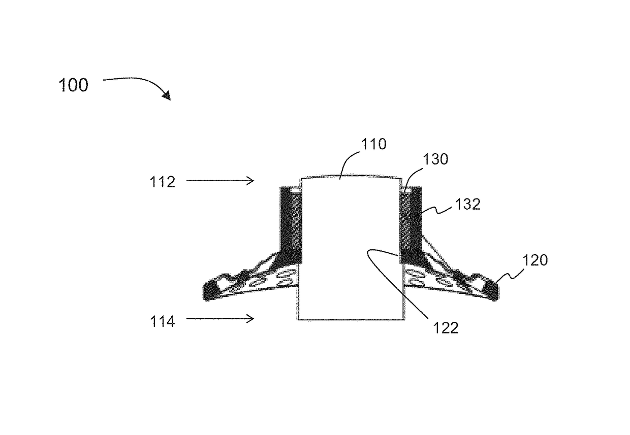
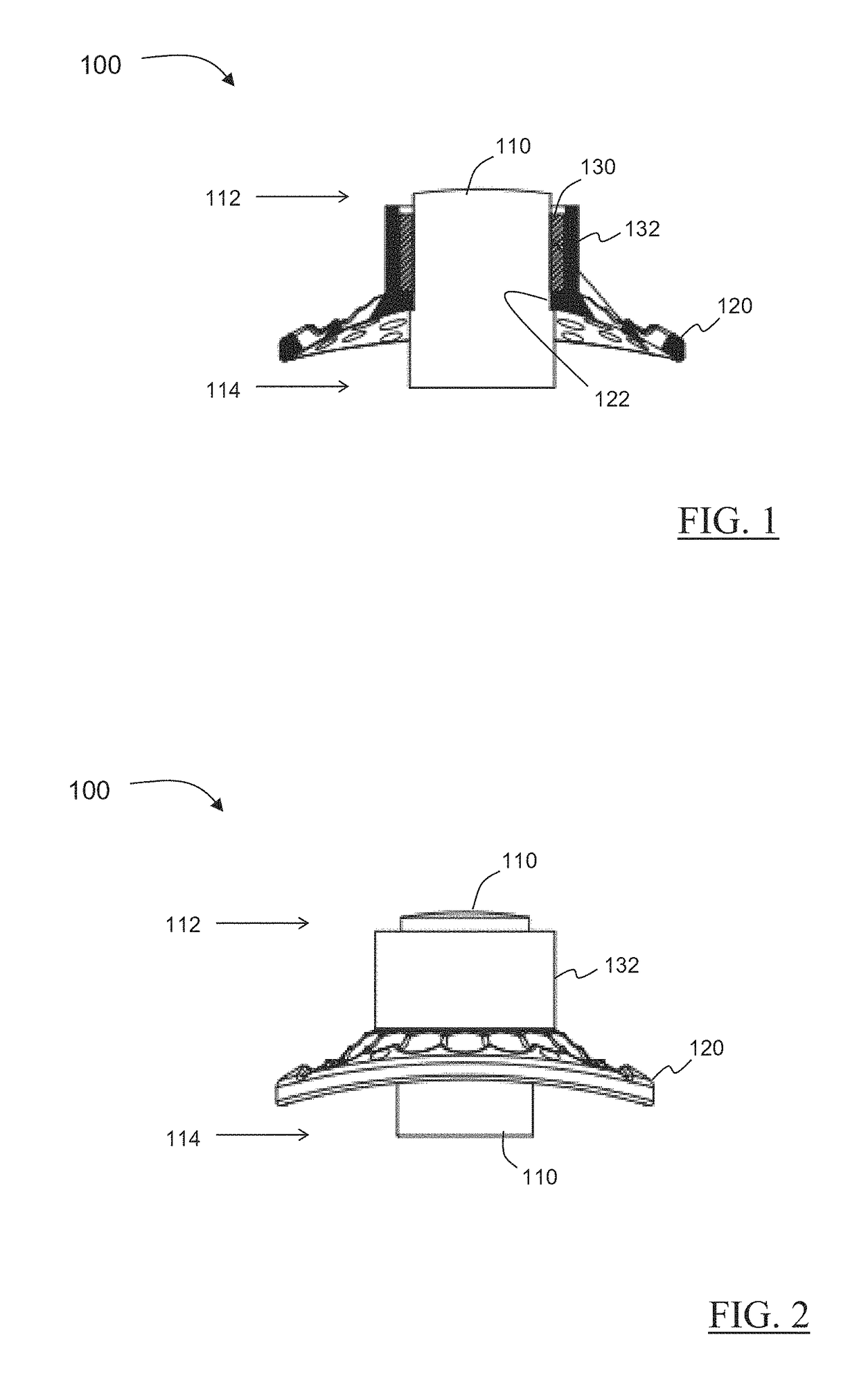
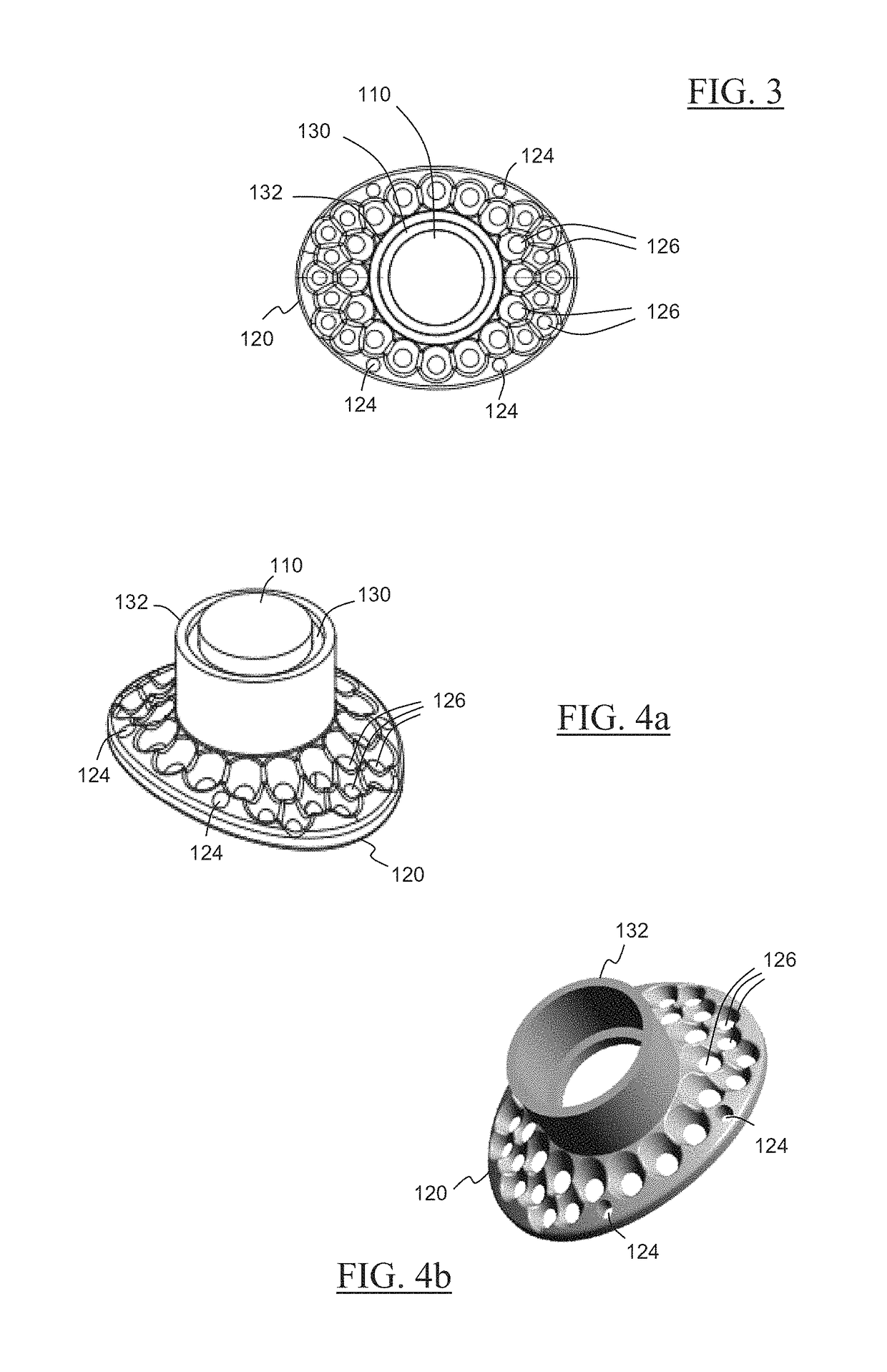
[0033]The present invention is directed to a novel keratoprosthesis and a system and method for implementing the same in repairing severe corneal damage. The present invention promotes the adhesion of soft tissue to the implanted keratoprosthesis to form a bioseal, prohibiting epithelial infiltration and extrusion that plagues currently available keratoprostheses. Moreover, the present keratoprosthesis produces no oral defects, and involves fewer ocular complications.

[0034]To begin, the keratoprosthesis of the present invention, shown throughout the Figures as 100, comprises an optic member 110 disposable in vision facilitating relation to the eye of a patient. As should be readily understood by those skilled in the art, the optic member is a cylinder or tube-like structure that houses a lens through which light passes in order for vision to occur. The optic member 110 used herein may be made from any appropriate and biologically compatible material, such as polymethylmethacrylate (...


PUM


Abstract

A keratoprosthesis and system and method of using same for corneal repair. The keratoprosthesis comprises a biocompatible support and an optic member disposed through a channel within the support. The support includes metal, preferably titanium, and treated, such as by sandblasting and / or acid etching, to create textured surfaces that promote soft tissue adhesion. A locking member interconnects the optic member and support. An outer surface of the locking member a collar extending from the support and disposed around the optic member is also metal, preferably titanium, and is similarly treated to promote soft tissue adhesion. A locking member interconnects the optic member and support. The system includes the keratoprosthesis positioned within an isolated soft tissue segment of a non-ocular tissue, such as buccal mucosa, placed on the anterior cornea. The method includes removing corneal epithelium, isolating and transplanting a segment of soft tissue to the de-epithelialized cornea, creating a receiving area in the soft tissue, positioning a keratoprosthesis relative to the receiving area anterior to the cornea, and securing the keratoprosthesis.

Description

CLAIM OF PRIORITY[0001]The present application is based on and a claim of priority is made under 35 U.S.C. Section 119(e) to a provisional patent application that is currently pending in the U.S. Patent and Trademark Office, namely, that having Ser. No. 60 / 696,937 and a filing date of Sep. 5, 2012, and which is incorporated herein by reference.STATEMENT OF GOVERNMENT SUPPORT[0002]This invention was made with government support under grant number W81XWH-09-1-0674 awarded by USARMY MEDCOM USAMRAA. The government has certain rights in the inventionBACKGROUND OF THE INVENTION[0003]1. Field of the Invention[0004]The present invention relates to corneal repair, and more in particular, to a novel keratoprosthesis that provides enhanced soft tissue adhesion and less extrusion, a system for repairing severely damaged cornea using such keratoprosthesis, and a method of implanting such keratoprosthesis to effect corneal repair.[0005]2. Description of the Related Art[0006]One of the greatest ch...

Claims


Application Information

Patent Timeline
Patent Type & Authority Patents(United States)
IPC IPC(8): A61F2/14
CPCA61F2/142
Inventor PAREL, JEAN-MARIESAWATARI, YOHPEREZ, VICTOR L.BERNAL, ANDRES
Owner UNIV OF MIAMI
Why Eureka
Try Eureka