
Human placental nutritive layer cell line and its use

A technology for trophoblast cells and human placenta, which can be applied in the field of cell lines to solve the problems of large individual differences and conflicting results.

Inactive Publication Date: 2008-07-23
INST OF ZOOLOGY CHINESE ACAD OF SCI
View PDF2 Cites 0 Cited by
  • Summary
  • Abstract
  • Description
  • Claims
  • Application Information

AI Technical Summary

Problems solved by technology

This is mainly due to the fact that human beings are used as research objects. Ethical issues have led to certain obstacles in obtaining human placenta tissue as a research material (especially in Western countries). Conflicting results

Method used


Image

  • Human placental nutritive layer cell line and its use
  • Human placental nutritive layer cell line and its use
  • Human placental nutritive layer cell line and its use

Examples


Embodiment 1

[0015] Example 1. Establishment of B6Tert-1 cell line

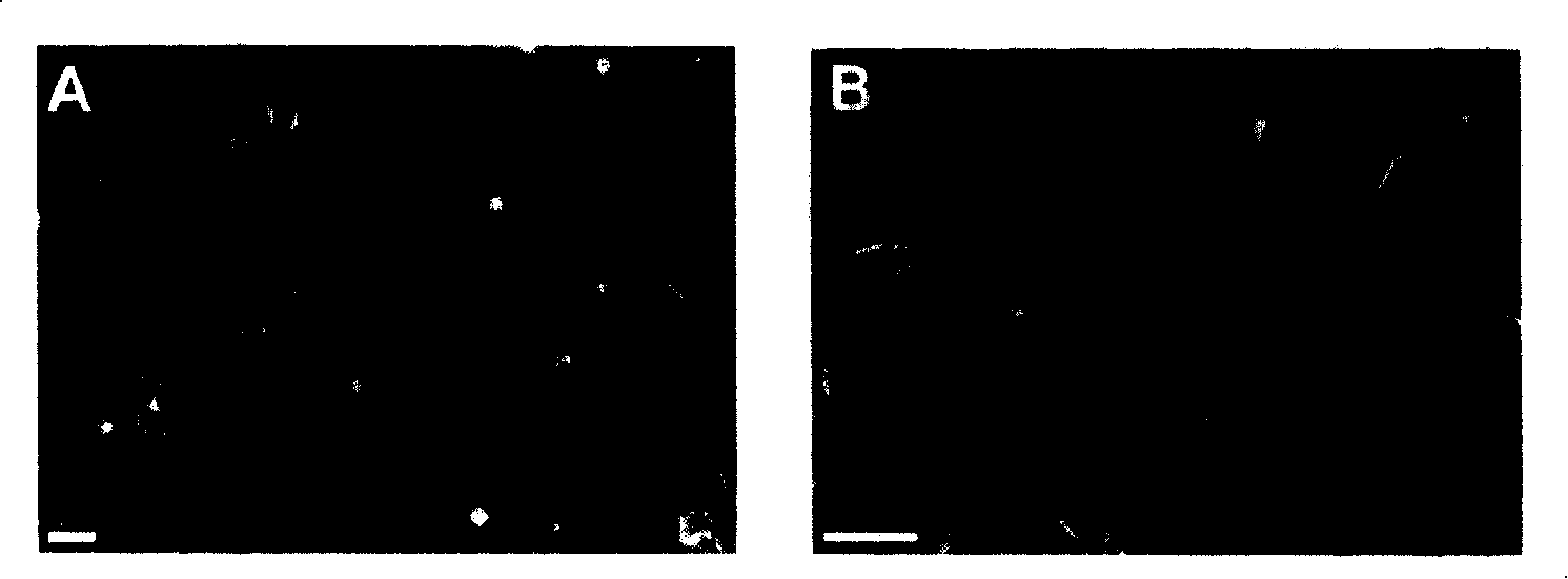
[0016] First, cytotrophoblast cells are isolated from the placenta of induced abortion at the age of 6-7 weeks of gestation (it can also be called villi at this time). Obtain 3-5 sterile villous tissues from the family planning clinic and bring them back to the laboratory in an ice bath; the following operations are all performed on a sterile operating table. Place the tissue in PBS buffer pre-cooled to 4°C to wash away the contaminated blood clots; transfer the tissue to fresh PBS buffer, separate the villi and basement membrane under a dissecting microscope and discard the basement membrane; collect the villi and Shred into 2-3mm 3 Transfer the small piece into a 50ml centrifuge tube, add 20ml of PBS buffer containing 0.2-0.3% trypsin (Sigma, USA), mix well and digest at 4-9°C for 30-50min; centrifuge (1000rpm, 5min) Discard the above solution, add 20ml of fresh FD culture medium and DNase (GIBCO, USA) at a final conc...

Embodiment 2

[0030] Example 2. Analysis of cell proliferation characteristics

[0031] 1) Cell proliferation rate

[0032] B6Tert-1(80PDs) cells according to 10 4 cells / dish inoculated on Cellmatrix-coated In the culture dish, three dishes were randomly selected every day from the 1st to the 11th day for cell counting, and the cell proliferation curve was drawn accordingly. The results showed that the cell doubling time of B6Tert-1 was 36 hours; the cells reached confluency (i.e., the cells were in close contact with no extra gaps in the culture dish) on day 9, at which point the cell proliferation slowed down and eventually stopped proliferating. This phenomenon, known as "contact inhibition" of cell growth, is one of the proliferative properties of normal cells.

[0033] 2) Regulation of cell proliferation by epidermal growth factor (EGF) and transforming growth factor (TGF) β:

[0034] B6Tert-1 cells were divided into 5×10 5 cells / dish inoculated on Cellmatrix-coated In the cult...

Embodiment 3

[0037] Example 3. Analysis of cell tumorigenicity (Tumorigenicity)

[0038] The B6Tert-1 cells grown to the 80 generation and the 210 generation were treated according to the 10 7 Cells / 100 μl were subcutaneously injected into nude mice respectively, and 3 nude mice were injected with each generation of cells. Tumor growth in nude mice was observed. After three months, no tumor formation was seen in the nude mice. It can be seen that B6Tert-1 cells are not tumorigenic.


PUM


Abstract

The invention relates to human placenta trophoblastic cell series which is conserved in China Committee for Culture Collection of Microorganisms on September 22th in 2005 with CGMCC No.1461 conserving number. It includes the following steps: separating the trophoblastic cell form the placenta tissue which is pregnant for 6-7 weeks; filtering the cells with multiplication capacity; processing subunit gene transfection by telomere enzyme to gain four long life cell strains. The cell series has normal multiplication capacity and biological chemical function, active cell growth, and reaches the 210 generation. Thus it can be used as target cell model for filtering antifertility drug and curing pregnant correlative disease medicine, provide ideal vitro model for further studying trophoblastic cell, lay important foundation for studying embryo implantation and pregnant physiology and pathological mechanisms.

Description

technical field [0001] The invention relates to a cell line, in particular to a human placental cytotrophoblast cell line and its application. Background technique [0002] During pregnancy, the placenta is an important endocrine organ that maintains the exchange of nutrients and substances between the mother and fetus, and is an important guarantee for the success of fetal development and pregnancy maintenance. The main component cells in the placenta are trophoblast cells, including mononuclear cytotrophoblast cells (CTB) with active proliferation ability and multinucleate syncytiotrophoblast cells (STB) that undertake the main endocrine function. STB is composed of CTB through the cell membrane formed by fusion. [0003] From the beginning to the end of pregnancy, most of the functions of the placenta are carried out by trophoblast cells. When the embryo is implanted, it relies on the identification, adhesion and invasion of the trophoblast cells and the endometrium to ...

Claims


Application Information

Patent Timeline
Patent Type & Authority Patents(China)
IPC IPC(8): C12N5/08A61K35/50C12R1/91C12N5/071
Inventor 王雁玲庄临之仇巍李玉侠李荣皓
Owner INST OF ZOOLOGY CHINESE ACAD OF SCI
Learn More