Method for detecting laser caused DNA damage

A DNA damage detection method technology, applied in the field of laser biology, to achieve the effect of easy promotion, simple method and accurate detection results

Inactive Publication Date: 2010-05-19
SHANDONG UNIV
View PDF0 Cites 0 Cited by
  • Summary
  • Abstract
  • Description
  • Claims
  • Application Information

AI Technical Summary

Problems solved by technology

[0004] To sum up, there are few biological detection methods for the biological effects of laser at present, and the damage of genetic material caused by laser is still only qualitative detection, especially at the DNA level, there is no effective and rapid detection method

Method used


Image

  • Method for detecting laser caused DNA damage
  • Method for detecting laser caused DNA damage
  • Method for detecting laser caused DNA damage

Examples


Embodiment 1

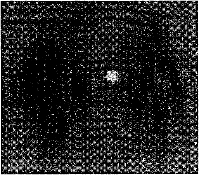
[0051] (1) Extraction of mouse splenocytes

[0052] A 4-week-old mouse was taken, killed by necking, and soaked in 75% alcohol for 10 minutes. Take out the spleen on a sterile operating table, peel off the fat tissue, and wash it with PBS buffer in a petri dish;

[0053] Place the cleaned mouse spleen on a 200-mesh stainless steel mesh and set it on a small beaker, cut the spleen into pieces with scissors, then grind the spleen with the piston core of the syringe, and wash the spleen with PBS buffer while grinding Tissue fragments, when the spleen tissue has basically been ground into a small beaker, remove the stainless steel mesh from the small beaker, pour the splenocyte suspension in the small beaker into a 10ml centrifuge tube, and wash the beaker 3 times with PBS buffer, Pour the cleaning solution into a centrifuge tube; centrifuge at 1200r / min for 6min, discard the supernatant, add 6ml of PBS buffer, blow off the cell pellet with a dropper to obtain a cell suspension; ce...

Embodiment 2

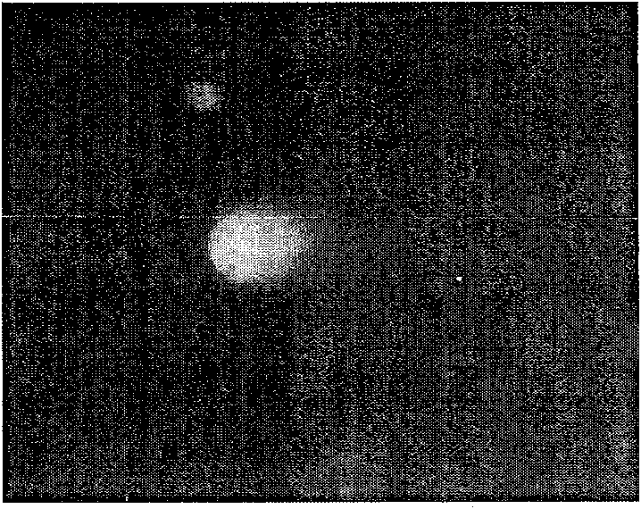
[0079] (1) Extraction of mouse splenocytes

[0080] A 3-week-old mouse was taken, killed by necking, and soaked in 75% alcohol for 5 minutes. Take out the spleen on a sterile operating table, peel off the fat tissue, and wash it with PBS buffer in a petri dish;

[0081] Place the cleaned mouse spleen on a 200-mesh stainless steel mesh and set it on a small beaker, cut the spleen into pieces with scissors, then grind the spleen with the piston core of the syringe, and wash the spleen with PBS buffer while grinding Tissue fragments, when the spleen tissue has basically been ground into a small beaker, remove the stainless steel mesh from the small beaker, pour the splenocyte suspension in the small beaker into a 10ml centrifuge tube, and wash the beaker 3 times with PBS buffer, Pour the cleaning solution into the centrifuge tube; centrifuge at 1000r / min for 8min, discard the supernatant, add 5ml of PBS buffer, blow off the cell pellet with a dropper to obtain a cell suspension;...

Embodiment 3

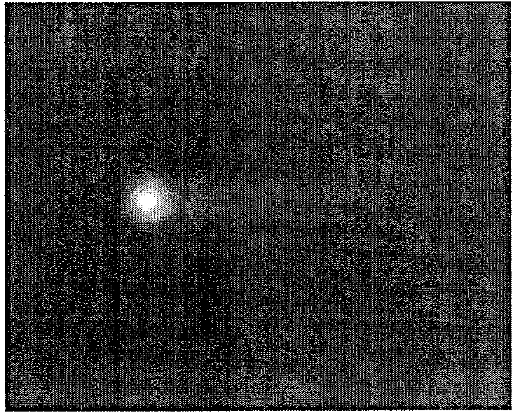
[0102] (1) Extraction of mouse splenocytes

[0103] A 6-week-old mouse was taken, killed by necking, and soaked in 75% alcohol for 15 minutes. Take out the spleen on a sterile operating table, peel off the fat tissue, and wash it with PBS buffer in a petri dish;

[0104] Place the cleaned mouse spleen on a 200-mesh stainless steel mesh and set it on a small beaker, cut the spleen into pieces with scissors, then grind the spleen with the piston core of the syringe, and wash the spleen with PBS buffer while grinding Tissue fragments, when the spleen tissue has basically been ground into a small beaker, remove the stainless steel mesh from the small beaker, pour the splenocyte suspension in the small beaker into a 10ml centrifuge tube, and wash the beaker 3 times with PBS buffer, Pour the cleaning solution into a centrifuge tube; centrifuge at 1500r / min for 5min, discard the supernatant, add 8ml of PBS buffer, blow off the cell pellet with a dropper to obtain a cell suspension; ...


PUM


Abstract

This invention discloses a method for detecting DNA damage caused by laser. The method comprises: radiating cells with laser, spreading gel onto prepared commnon glass slide with improved single-cellgel electrophoresis method, performing electrophoresis, coloring, observing the result under fluorescence microscope, analyzing the images with CASP software to determine DNA damage, and calssifying the damage. The method can easily, rapidly and quantitatively determine DNA damage caused by laser on molecular level, and does not need any specific apparatus. The observation can even be performed under inverted fluorescence microscope. Besides, the experiment only needs a small quantity of cells, and is nontoxic to experimenters.

Description

technical field [0001] The invention relates to a cell detection method, in particular to a laser-induced DNA damage detection method, which belongs to the field of laser biology. Background technique [0002] "Laser" is an abbreviation for stimulated radiation amplification of light, which achieves light amplification by stimulated emission of radiation. Compared with ordinary light, laser light has good directionality, concentrated energy, relatively single wavelength, and good coherence. Therefore, when it irradiates biological tissues or cells and interacts with biological substances, in addition to the biological effects caused by ordinary light of the same wavelength, it can also cause many unique biological effects, such as light effects, electromagnetic field effects, thermal effects and pressure effects. . In 1960, Maiman of the United States made the world's first ruby ​​laser. Since then, the development of laser has advanced by leaps and bounds, and great progr...

Claims


Application Information

Patent Timeline
Patent Type & Authority Patents(China)
IPC IPC(8): C12Q1/68G01N33/48G01N27/447G01N21/64
Inventor 时永香张楠吴克良张乐白增亮张少军
Owner SHANDONG UNIV
Why Eureka
Try Eureka