
Pharmaceutical in vivo dynamics characteristic-nondestructive in situ monitoring system and monitoring method

A monitoring system and drug technology, applied in pharmaceutical formulations, pharmaceutical sciences, preparations for in vivo experiments, etc., to achieve the effect of reducing background interference

Inactive Publication Date: 2007-08-08
CHINA PHARM UNIV
View PDF7 Cites 8 Cited by
  • Summary
  • Abstract
  • Description
  • Claims
  • Application Information

AI Technical Summary

Problems solved by technology

However, the premise of this method is that the peptide is immobilized on the resin, and the excess dye can be eluted with organic reagents. It is not suitable for the case where the protein or peptide is in solution.

Method used

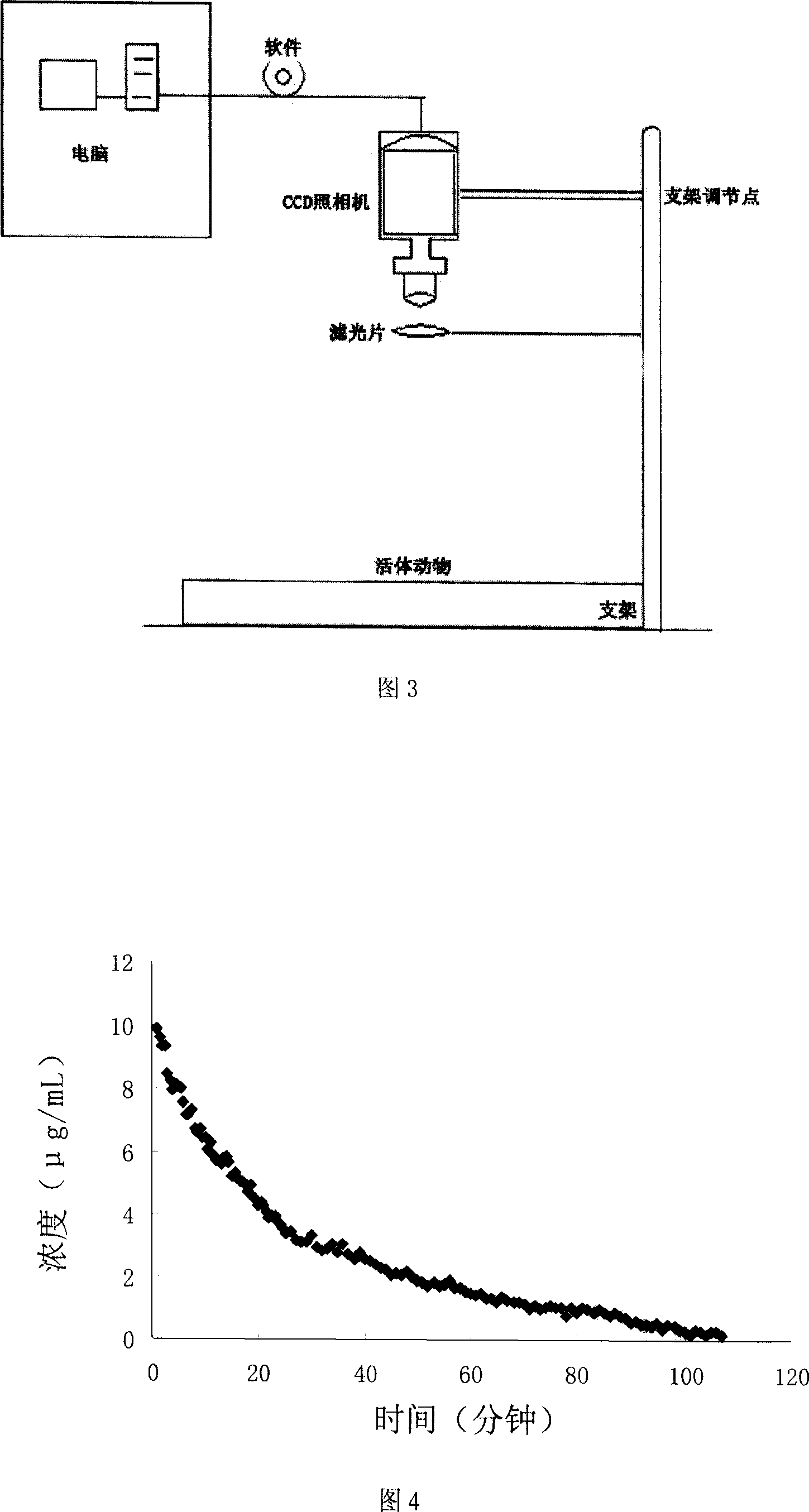

Image

  • Pharmaceutical in vivo dynamics characteristic-nondestructive in situ monitoring system and monitoring method
  • Pharmaceutical in vivo dynamics characteristic-nondestructive in situ monitoring system and monitoring method
  • Pharmaceutical in vivo dynamics characteristic-nondestructive in situ monitoring system and monitoring method

Examples


Embodiment 1

[0043] Synthetic method of cypate-labeled insulin

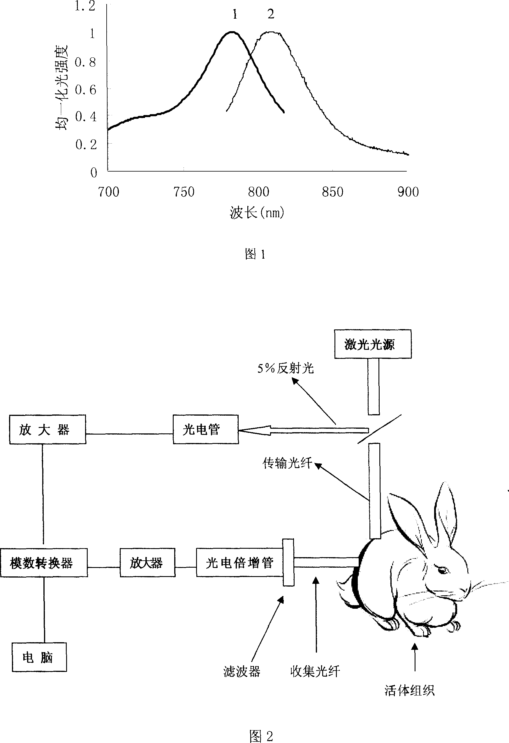
[0044] Dissolve 1mg cypate (see Figure 1 for the spectrum) in anhydrous DMSO, add 0.4mgHBTU and 0.3mgHOBT to it, vortex and mix well in the dark, then slowly add 4ml 1.25mg / ml of the activation solution dropwise while shaking Insulin solution (pH8.5), stirred and reacted at 4 degrees for 6 hours. After the reaction solution was mixed, it was dialyzed in PBS buffer solution (pH7.4) for two days, and the dialysate was changed every 4 hours. The resulting solution was lyophilized and stored in a -20°C refrigerator. The calculated labeling ratio of cypate to insulin was 0.57:1.

Embodiment 2

[0046] Synthetic method of cypate-labeled lysozyme

[0047] 1 mg of cypate was dissolved in anhydrous DMSO, 0.4 mg of DCC was added thereto, after vortex mixing, the reaction was shaken for 30 minutes. Add 0.3 mg HOBT into the solution, stir for 2 hours, and centrifuge to remove the supernatant to obtain the dye activation solution. The activation solution was slowly dropped into 4ml of 1.25mg / ml lysozyme solution (pH9.2) while shaking, and stirred and reacted at 4°C for 6 hours. After the reaction solution was mixed, it was dialyzed in PBS buffer solution (pH7.4) for two days, and the dialysate was changed every 4 hours. The resulting solution was lyophilized and stored in a -20°C refrigerator. The calculated labeling ratio of dye cypate to lysozyme was 1.94:1.

Embodiment 3

[0049] Synthetic method of cypate-marked recombinant L-asparaginase

[0050] Dissolve 1 mg of cypate in anhydrous DMSO, add 0.4 mg of HBTU and 0.3 mg of HOBT to it, vortex and mix well in the dark, then slowly add 4 ml of 1.25 mg / ml L-asparagine dropwise to the activation solution while shaking In the enzyme solution (pH8.5), the reaction was stirred at 4 degrees for 6 hours. After the reaction solution was mixed, it was dialyzed in PBS buffer solution (pH7.4) for two days, and the dialysate was changed every 4 hours. The resulting solution was lyophilized and stored in a -20°C refrigerator. The labeling ratio of cypate to asparaginase was calculated to be 33:1.


PUM

PropertyMeasurementUnit
Wavelengthaaaaaaaaaa

Abstract

The invention relates to a system used in the internal dynamic nondestructive online checking of protein polypeptide drug, and relative checking method. The inventive system comprises a fluorescence probe and a detecting system, while the detecting system is formed by a near-infrared light source, a transmission optical fiber, a receiving optical fiber, a near-infrared high-pass filter, and a near-infrared detector. The laser of light source and the fluorescence light received by the detector can be transmitted by the optical fiber; the near-infrared band-pass or high-pass filter is at the front of detector. The invention can avoid sampling timely in the test, avoid killing animals in the organism distribution test, to and avoid the following sample treatment.

Description

technical field [0001] The invention relates to the field of in vivo detection of drugs, in particular to a system and a detection method for non-destructive in situ detection of in vivo kinetic properties of protein and polypeptide drugs. Background technique [0002] Protein peptide drugs have strong physiological activity and high therapeutic index, and play an important role in maintaining the normal function of the body. The study of drug kinetics in vivo is an essential part of any drug development process. At present, the detection methods of protein and polypeptide drugs in vivo can be roughly summarized into four types: immunological detection method, isotope labeling and tracing method, physical and chemical analysis technology and biological assay method. Except for the isotope tracer method, the other three detection methods cannot realize on-site real-time monitoring, and need to go through procedures such as regular sampling, sample processing, and analytical ...

Claims


Application Information

Patent Timeline
IPC IPC(8): A61B5/00A61K49/00A61D99/00
Inventor 顾月清钱慧敏方春生钱志余
Owner CHINA PHARM UNIV
Learn More