Method for enhancing detection reagent sensitivity by using colloidal gold or latex granule as marker
A technology of latex particles and colloidal metals, applied in measurement devices, instruments, scientific instruments, etc., can solve the problems of pathogens affecting the application of reagents and low detection sensitivity, and achieve the effect of improving specificity and increasing sensitivity
- Summary
- Abstract
- Description
- Claims
- Application Information
AI Technical Summary
Problems solved by technology
Method used
Image
Examples
Embodiment 1
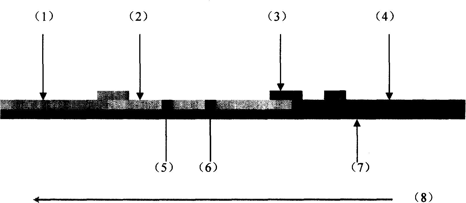
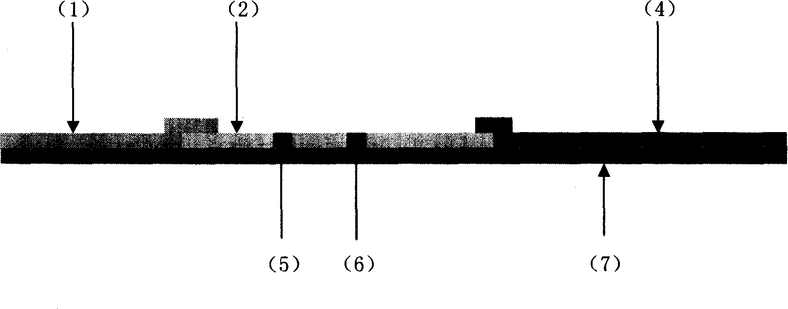
[0053] Example 1: Respiratory syncytial virus latex method detection kit
[0054] (1) Purchase latex particles with suitable particle size.
[0055] (2) Marking of latex particles
[0056] Label according to the steps in the instruction manual of the latex particles, and label the anti-respiratory syncytial virus monoclonal antibody on the latex particles. Centrifuge and purify the labeled latex particles at an appropriate speed, carefully suck off the supernatant, and suspend the precipitate with preservation solution to make latex particle-antibody conjugates.
[0057](3) Drying of latex particle-antibody conjugate
[0058] Take the latex particle-antibody conjugate solution, dilute it to an appropriate concentration with the working solution, and distribute it into 2 ml vials or small tubes. Freeze-dry overnight in a freeze dryer until completely dry. After sealing, keep airtight and dry.
[0059] (4) Antibody kit for nitrocellulose membrane
[0060] ① Dilute the anti...
Embodiment 2
[0083] Embodiment 2: A and B influenza virus colloidal gold detection kits
[0084] (1) Preparation of colloidal gold Colloidal gold particles of 20 to 50 nm were prepared by chemical reduction hair or other suitable methods.
[0085] (2) Marking of colloidal gold
[0086] The colloidal gold solution prepared in step (1) was treated with 0.2M K 2 CO 3 Adjust to a suitable pH value. Add the anti-influenza A virus monoclonal antibody into the above solution in an appropriate labeled amount, and stir at room temperature. Add stabilizers such as BSA and PEG of appropriate concentration, and stir at room temperature. Centrifuge at an appropriate speed until the supernatant is basically colorless, carefully suck off the supernatant, and suspend the precipitate with colloidal gold preservation solution.
[0087] The colloidal gold solution prepared in step (1) was treated with 0.2M K 2 CO 3 Adjust to a suitable pH value. Add the anti-influenza B virus monoclonal antibody into...
Embodiment 3
[0117] Embodiment 3: Treponema pallidum colloidal selenium detection kit
[0118] (1) Purchase or prepare colloidal selenium solution with suitable particle size.
[0119] (2) Labeling of colloidal selenium
[0120] Follow the appropriate steps for labeling and label the anti-Treponema pallidum monoclonal antibody onto the colloidal selenium particles. Centrifuge and purify the labeled latex particles at an appropriate speed, carefully suck off the supernatant, and suspend the precipitate with the preservation solution to prepare the colloidal selenium-antibody conjugate.
[0121] (3) drying of colloidal selenium-antibody conjugate
[0122] Take the colloidal selenium-antibody conjugate solution and dilute it with working solution to an appropriate concentration.
[0123] Spread the glass fiber or PE composite membrane on a glass plate, and evenly drop the diluted colloidal selenium-antibody conjugate solution on it. Freeze-dry overnight in a freeze dryer until completely ...
PUM
 Abstract
Description
Claims
Application Information
 - R&D Engineer
- R&D Manager
- IP Professional
- Industry Leading Data Capabilities
- Powerful AI technology
- Patent DNA Extraction
Browse by: Latest US Patents, China's latest patents, Technical Efficacy Thesaurus, Application Domain, Technology Topic, Popular Technical Reports.
© 2024 PatSnap. All rights reserved.Legal|Privacy policy|Modern Slavery Act Transparency Statement|Sitemap|About US| Contact US: help@patsnap.com


