Preparation, storage and use of umbilical cord and placenta mesenchymal stem cell for clinical therapy

A kind of stem cell and stem cell technology, applied in the field of umbilical cord placenta stem cells, can solve the problems of umbilical cord pollution, cross-contamination of other specimens, cross-contamination of liquid phase, etc., to achieve clinical application guarantee, save storage space, and avoid pollution effects

Inactive Publication Date: 2008-11-05
TIANJIN HUANYUSNGQIAO BUSINESS INFORMATION CONSULTING
0 Cites 19 Cited by

AI-Extracted Technical Summary

Problems solved by technology

Many normal delivery umbilical cords are not processed well, making them unusable for stem cell storage
[0007] 2Because some umbilical cords are contaminated by pathogens, some umbilical cords obtained during the culture process, bacteria, molds, etc. reproduce, causing pollution to other cells and other individual storage
[0008] 3 What is more serious is that at present, the method of immersion in liquid nitrogen is often used in general storage, which will increase the risk of cross-contamination between the stored cells through the liquid nitrogen liquid phase
[0009] 4 When using a plastic storage tube with a cover, the chance of cross-contamination of other specimens is also greatly increased due to leakage from the cover of the tube
[0010] The umbilical...

Abstract

The invention discloses the preparation, the storage and an application of a stem cell used for the clinical care. The establishment of the invention comprises the following steps that: 1) the decontamination treatment of a placenta and a umbilical cord is performed; a) medical iodine and alcohol or other disinfector are adopted to process the surfaces of the placenta and the umbilical cord; b) the outer surfaces of the placenta and the umbilical cord are removed by an instrument or manually; c) the loss of the bacterium and the stem cell of the placenta and the umbilical cord tissue; 2) the preparation and culture without bacteria of the mesenchyma stem cell; a) the tissue is processed manually or automatically without bacteria; b) the tissue is grinded into the smallest particles without bacteria; c) the tissue in the 2b item of the enzyme treatment is prepared; d) the mixture is washed and separated centrifugally to collect the cell; e) the cell is cultured; 3) the umbilical cord mesenchyma stem cell is stored safely; a) the cell is collected into a storage pipe or a bag in a sterile way; b) a sealing cover is added on the storage pipe; c) the double layer of the high quality plastic film is covered and each pipe is sealed by a pumping method; d) the cell is stored inside a fluid phase of a steam type liquid nitrogen storage tank, or the safe injection fluid is prepared.

Application Domain

Technology Topic

Culture cellInstrumentation +13

Image

  • Preparation, storage and use of umbilical cord and placenta mesenchymal stem cell for clinical therapy
  • Preparation, storage and use of umbilical cord and placenta mesenchymal stem cell for clinical therapy

Examples

  • Experimental program(3)

Example Embodiment

[0035] Example 1:
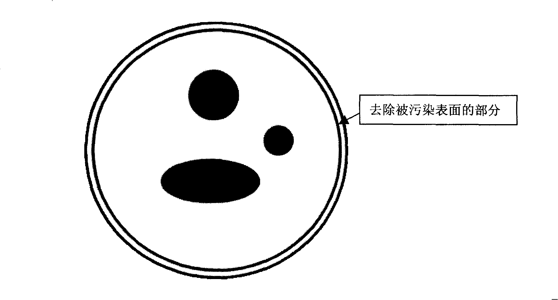
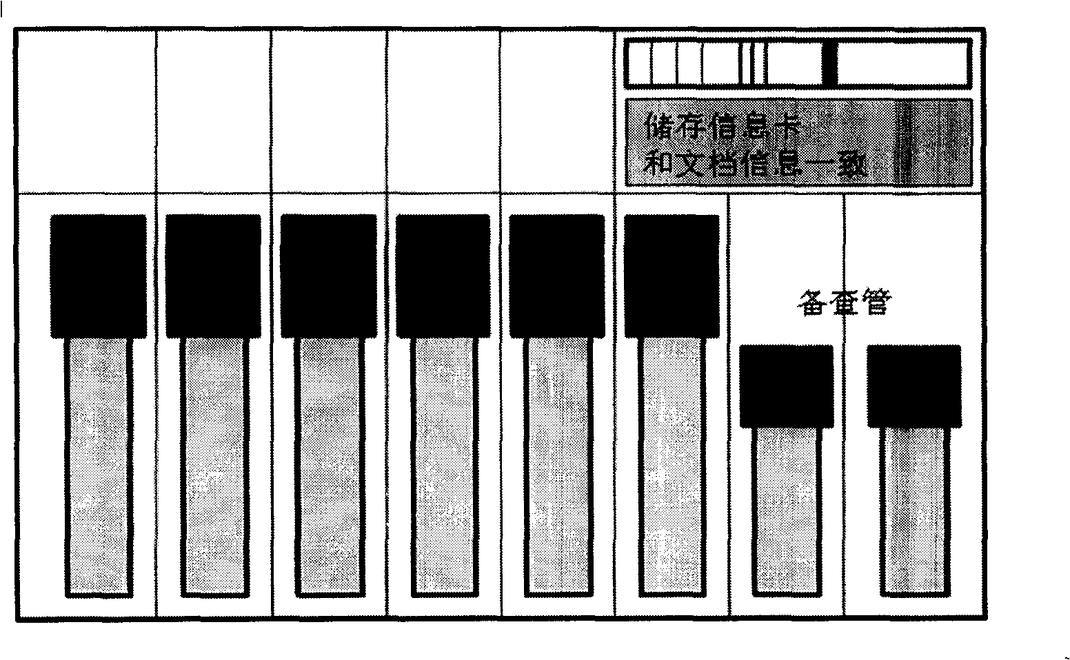
[0036] Normal vaginal delivery placenta, decontamination of umbilical cord. Flush the placenta and umbilical cord with sterile saline to remove visible dirt and residual blood. Use 2% medical iodine for 30 seconds, then deiodine with 75% medical alcohol. The outer surface of the umbilical cord placenta was separated by hand about 0.5 mm. The outer surface does not touch the sterile inner tissue during removal. The stored individuals are routinely tested for tissue typing (HLA), biology, and pathogenic microorganisms. At the same time, we will establish databases for clinical umbilical cords and placental mesenchymal stem cells, and prepare data cards and barcodes that are consistent with the various types of cells stored, personal data databases, and various reports. Preparation and culture of mesenchymal stem cells.
[0037] Umbilical cord or placental tissue is processed to less than 1 mm in size using sterile manual, or automated methods. Centrifuge at 250g for 5 minutes at room temperature, discard the supernatant, collect the cells, and wash with DMEM. Centrifuge and wash again. Conventional collagenase, trypsin treated tissue. Routinely collect and store the first generation of mesenchymal stem cells and collect the second and third generation mesenchymal stem cells after culture. Dulbecco's modified eagle's medium (DMEM, Gibco, USA) and 10% autoserum culture medium were used for culture.
[0038] Umbilical cord mesenchymal stem cells were aseptically collected as needed in sterile 5ml cell storage tubes. 5mL tube, (430663), medium, 2ml as the tube to be checked. The storage tube containing the stem cells is capped. The cell storage tubes after capping are further covered with high-quality polyester film, double-layer film and each tube is sealed by evacuation sealing method. Each depositor has its own sealed storage system. The respective systems contain barcodes of seals and depositor information consistent with computer and paper file archives. These individual stem cell storage systems are stored in the vapor phase of MVE high-efficiency steam liquid nitrogen tank Vapor (MVE 815P-190, MVE 1536P-190, MVE 1879P-190) at -190 degrees Celsius. For the sake of safety and actual clinical needs, the inspection management is divided into two levels of storage and inspection. Store one copy in a -80°C freezer and the other in the airtight storage system described above.

Example Embodiment

[0039] Example 2:
[0040]Normal vaginal delivery placenta, decontamination of umbilical cord. Flush the placenta and umbilical cord with sterile saline to remove visible dirt and residual blood. Use 2% medical iodine and 75% medical alcohol to treat the surface of the umbilical cord and placenta. According to the situation, about 0.5mm of the outer surface of the umbilical cord placenta was removed manually and with special instruments. The outer surface does not touch the sterile inner tissue during removal. The stored individuals are routinely tested for tissue matching (HLA), biology, and pathogenic microorganisms. At the same time, we will establish databases for clinical umbilical cords and placental mesenchymal stem cells, and prepare data cards and barcodes that are consistent with the various types of cells stored, personal data databases, and various reports. Preparation and culture of mesenchymal stem cells. Allows automated methods to process umbilical cord or placental tissue to less than 1mm size. The tissue is further processed to smaller particles using a sterile tissue grinding facility. Centrifuge at 250g for 5 minutes at room temperature, discard the supernatant, collect the cells, and wash with DMEM. Centrifuge and wash again. Conventional collagenase, trypsin treated tissue. Routinely collect and store the first generation of mesenchymal stem cells and collect the second and third generation mesenchymal stem cells after culture. Dulbecco's modified eagle's medium (DMEM, Gibco, USA) and serum-free medium were used for culture.
[0041] Safe storage of umbilical cord mesenchymal stem cells in frozen bags for further use The bags were covered with FEP film and sealed with evacuation seal. Each depositor has its own sealed storage system. The respective systems contain barcodes of seals and depositor information consistent with computer and paper file archives. These individual stem cell storage systems are stored in the vapor phase portion of vaporized liquid nitrogen tanks. For the sake of safety and actual clinical needs, the inspection management is divided into two levels of storage and inspection. Store one copy in a -80°C freezer and the other in the airtight storage system described above. All the cells treated by the above methods were stored in the vapor phase of MVE high-efficiency steam liquid nitrogen tank Vapor (MVE 815P-190, MVE 1536P-190, MVE 1879P-190) at -190 degrees Celsius.

Example Embodiment

[0042] Example 3:
[0043] The placenta and umbilical cord were flushed with sterile saline containing penicillin and streptomycin to remove residual blood. With medical H 2 o 2 Quickly process placenta, umbilical cord surface. According to the situation, the outer surface of the umbilical cord placenta of about 0.5 mm is removed by manual separation or special equipment. The outer surface does not touch the sterile inner tissue during removal. The stored individuals are routinely tested for tissue typing (HLA), biology, and pathogenic microorganisms. At the same time, we will establish databases for clinical umbilical cords and placental mesenchymal stem cells, and prepare data cards and barcodes that are consistent with the various types of cells stored, personal data databases, and various reports.
[0044] Umbilical cord or placental tissue is processed to less than 1 mm in size using sterile manual, or automated methods. Tissue can further be processed to smaller particles using sterile tissue grinding facilities. Centrifuge at room temperature at 250g for 5 minutes, discard the supernatant, collect the cells, and wash with DMEM with penicillin and streptomycin. Centrifuge and wash again. Conventional collagenase, trypsin treated tissue. Routinely collect and store the first generation of mesenchymal stem cells and collect the second and third generation mesenchymal stem cells after culture. Dulbecco's modified eagle's medium (DMEM, Gibco, USA), normal human AB serum, and 1% glutamine (Gibco, USA) were used for culture. Umbilical cord mesenchymal stem cells were aseptically collected as needed in sterile cryogenic cryovials. The storage tube containing the stem cells is capped. The capped cell storage tube is further covered with high-quality polyester film, EVO copolymer film, Cover each tube with FEP film or other double-layer film suitable for -210°C and seal it with evacuation sealing. Each depositor has its own sealed storage system. The respective systems contain barcodes of seals and depositor information consistent with computer and paper file archives. These individual stem cell storage systems are stored in the vapor phase portion of vaporized liquid nitrogen tanks. For the sake of safety and actual clinical needs, the inspection management is divided into two levels of storage and inspection. Store one in a -80°C freezer and the other in an airtight storage system. All the cells treated by the above methods are stored in the vapor phase of the MVE high-efficiency steam liquid nitrogen tank Vapor (MVE 815P-190, MVE 1536P-190, MVE 1879P-190) at -190 degrees Celsius, or -190 degrees Celsius from other manufacturers In the vapor phase of the vapor-type liquid nitrogen storage tank.
[0045] The stem cells processed by the above method, and the stem cell bank and storage method thereof can avoid cross-contamination of bacteria, mold and other pathogens, other human cells, DNA, RNA, etc. caused by liquid phase immersion. Pollution to stem cells of other depositors caused by leakage of the used tube cap can also be avoided, so that the safety and future clinical application of the stored stem cells can be guaranteed. At the same time, it saves a lot of storage space. Thus reducing costs.

PUM

no PUM

Description & Claims & Application Information

We can also present the details of the Description, Claims and Application information to help users get a comprehensive understanding of the technical details of the patent, such as background art, summary of invention, brief description of drawings, description of embodiments, and other original content. On the other hand, users can also determine the specific scope of protection of the technology through the list of claims; as well as understand the changes in the life cycle of the technology with the presentation of the patent timeline. Login to view more.

Similar technology patents

Classification and recommendation of technical efficacy words

Why Eureka
Try Eureka