Tissue engineering trachea implant and construction method and use

A technology of tissue engineering and grafting, which is applied in the field of medicine and biomedical engineering, and can solve the problems of mixing mouse cells, failing to show the superiority of tissue engineering technology, and insufficient cartilage support strength, etc.

Inactive Publication Date: 2008-12-17
SHANGHAI TISSUE ENG LIFE SCI
View PDF0 Cites 4 Cited by
  • Summary
  • Abstract
  • Description
  • Claims
  • Application Information

AI Technical Summary

Problems solved by technology

[0008] 1) The cartilage support strength of the tissue engineered trachea constructed purely in vitro is insufficient, making it difficult to maintain the ventilation function;
[0009] 2) Although the simple construction in vivo is helpful to improve the mechanical strength of cartilage, direct placement in the body will cause a strong inflammatory response due to the acidic degradation products of commonly used PGA materials in animals, and it is difficult to form a good tissue engineering trachea;
[0010] 3) Some studies choose to construct in nude mice first to reduce the above-mentioned inflammatory response, but the mouse-derived cells are mixed into the tissue-engineered trachea, and then transplanted into experimental animals with sound immunity will cause heterogeneous immune rejection;
[0011] 4) In the pr

Method used


Image

  • Tissue engineering trachea implant and construction method and use
  • Tissue engineering trachea implant and construction method and use
  • Tissue engineering trachea implant and construction method and use

Examples


Embodiment 1

[0107] Cell Isolation and Culture

[0108] Sterilize appropriate amount of cartilage, cut into 1mm pieces 3 Cartilage particles of large or small sizes were first digested with 0.05% trypsin solution at 37°C for 30 minutes with shaking, then washed with PBS and added with 0.25% type II collagenase solution (Sigma Company), and digested with constant temperature shaking at 37°C for 7-8 hours. The digestive solution was filtered through a 100 μm filter, and after centrifugation, the cell pellet was resuspended in chondrocyte culture medium, counted, inoculated into a 10mm culture dish (DownCorning Company) at a cell density of 1.0×10⁴ / cm2, and chondrocyte culture medium was added; the cells were incubated at 37°C , 5% CO2, cultured under saturated humidity conditions, the first medium was changed after 3 days, and the medium was changed every other day thereafter. The formula of chondrocyte culture medium is: DMEM (Dulbecco'smodified Eagle's medium) culture medium (Gibco Compan...

Embodiment 2

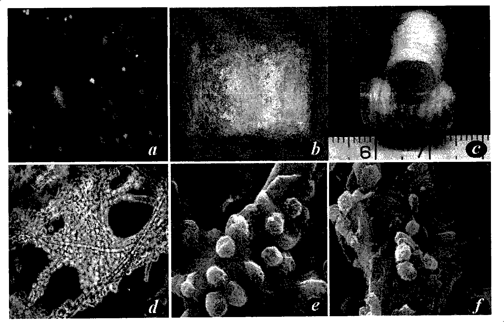
[0111] Scaffold material preparation

[0112] Take 60 mg of non-woven polyglycolic acid fiber (polyglycolic acid, PGA, self-developed), and spirally wind it on a silicone tube with a length of 1.5 cm and a diameter of 6 mm to form a cylindrical scaffold material ( figure 1 ), both ends were sutured and fixed with absorbable sutures, soaked in 75% alcohol for 60 minutes for disinfection, rinsed with PBS twice and then set aside.

Embodiment 3

[0114] Cell-material complex formation and in vitro culture

[0115] Chondrocytes adjusted cell concentration to 60×10 6 / ml, take 1ml of cell suspension and inoculate it evenly on the spiral material, place it at 37°C, 5% CO2, and saturated humidity for 4 hours, then transfer it into a 50ml polypropylene centrifuge tube (BD Biosciences), add 30ml of chondrocyte culture medium, and change it every other day The culture was continued for 2 weeks under the above conditions.


PUM

PropertyMeasurementUnit
Lengthaaaaaaaaaa

Abstract

The invention provides a trachea implant in tissue engineering. Chondroblast is combined with degradable biomaterial and then cultured and constructed jointly in vitro and in vivo, and has the blood supply connection with cultured tissues in vivo. The trachea implant in tissue engineering of the invention can effectively repair the segmental defect of the trachea. The invention further provides a construction method and a use method thereof.

Description

technical field [0001] The present invention relates to the fields of medicine and biomedical engineering, and more particularly relates to a tissue engineered trachea graft and its construction method and application. Background technique [0002] Tracheal defects caused by various reasons (congenital malformation, trauma, tumor, etc.) are common and life-threatening. The mortality rate of long-segment tracheal stenosis can be as high as 77%, and the mortality rate of long-segment tracheal atresia is basically 100%. Long-segment tracheal defects cannot be anastomosed directly, so tracheal substitutes are required for repair. Currently, there are three types of tracheal transplantation substitutes commonly used clinically, but each has its own shortcomings, as shown in Table 1. Therefore, there is still a lack of ideal tracheal substitutes, and the repair of long-segment tracheal defects has thus become a clinical problem. [0003] The development of tissue engineering pro...

Claims


Application Information

Patent Timeline
IPC IPC(8): A61L29/12A61L29/14A61F2/04
Inventor 周广东曹谊林罗旭松刘伟崔磊
Owner SHANGHAI TISSUE ENG LIFE SCI
Why Eureka
Try Eureka