Implantation type artificial hepar

An implantable and artificial technology, applied in the fields of biomedicine, regenerative medicine, and clinical medicine, can solve the problems of lack of biological correspondence and the inability of implanted cells to eventually replace organ function, so as to promote angiogenesis, promote long-term survival and function. , the effect of strong tissue regeneration

Active Publication Date: 2009-05-13
ZHEJIANG UNIV
View PDF0 Cites 13 Cited by
  • Summary
  • Abstract
  • Description
  • Claims
  • Application Information

AI Technical Summary

Problems solved by technology

However, the currently commonly used tissue engineering scaffolds are porous materials with basically the same functions. The chemical properties and basic physical properties of the whole material are roughly uniform, and there is no sp

Method used


Image

  • Implantation type artificial hepar
  • Implantation type artificial hepar
  • Implantation type artificial hepar

Examples


specific Embodiment 1

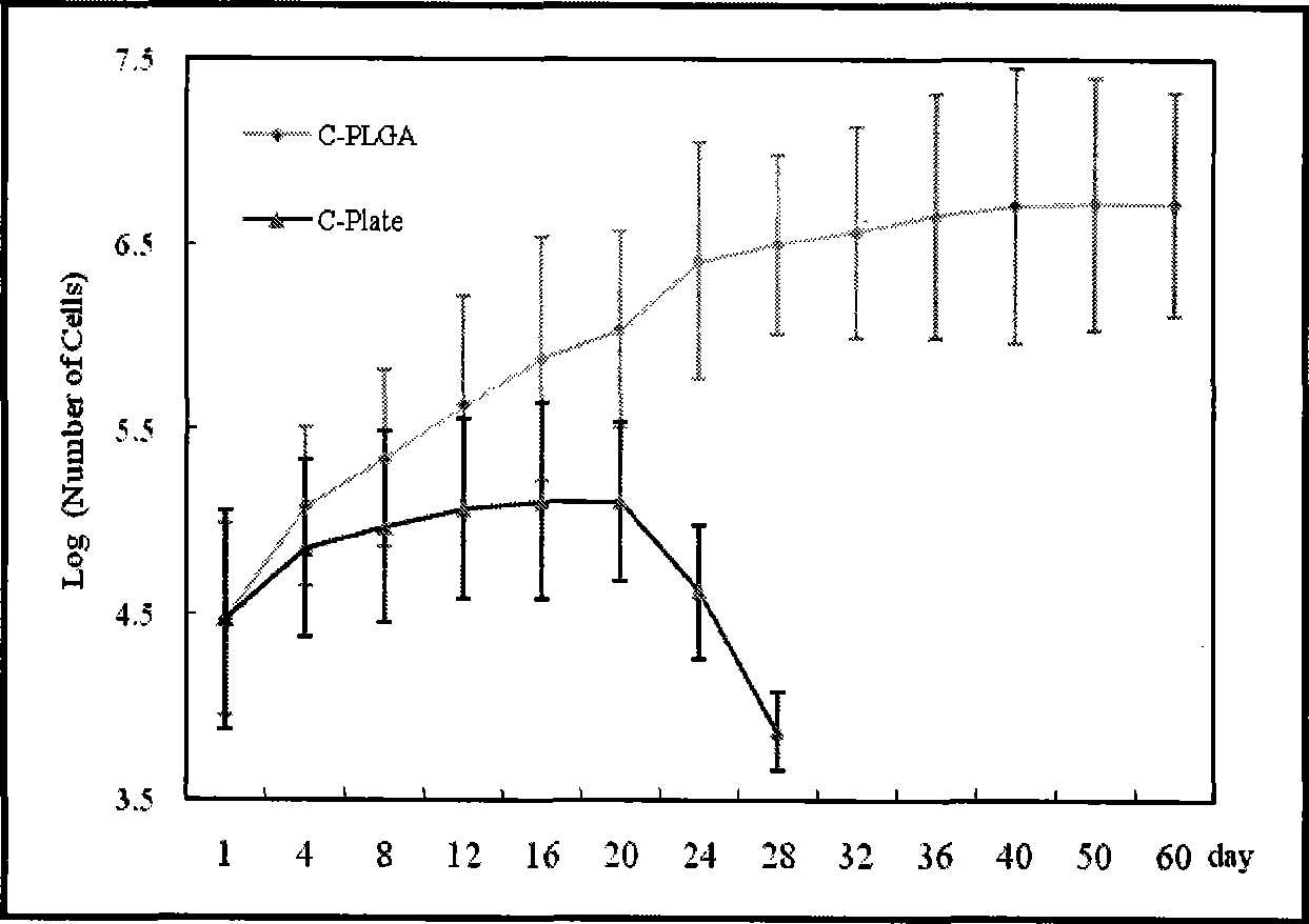
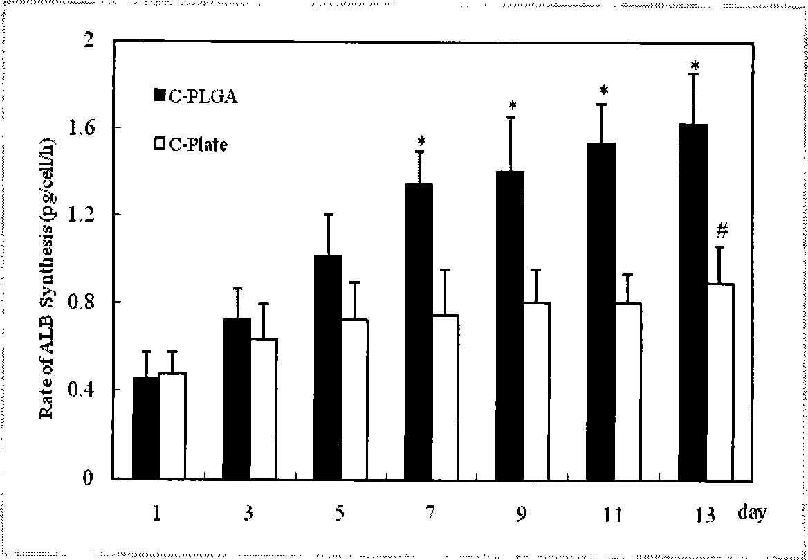
[0025] Specific Example 1 (Comparison of Implantable Artificial Liver and Monolayer Adhesive Culture Rat Hepatocytes)

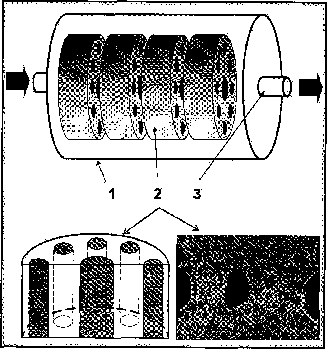
[0026] The polyglycolic acid or polylactic acid glycolic acid envelope 1 is 15 mm long, and the material of the three-dimensional porous support 2 is PLGA, and the molecular weight of PLGA is 70,000 units. The porous scaffold 2 has a diameter of 9mm, a thickness of 3mm, and a pore diameter of 100-200nm, with a specific lumen (the lumen structure is a circular lumen with a pore diameter of 200-500nm, which is directly laser-drilled in the three-dimensional stent, No other materials, mainly used to provide endothelial cell growth and induce microangiogenesis.) 6-8, with a pore diameter of 400nm, stacked with 2 sets of 4 porous scaffolds. Polyglycolic acid or polylactic-glycolic acid tube seal 3 diameter 2 mm.

[0027] Rat primary hepatocytes obtained by conventional separation, with 10 7 The cells / ml density was inoculated into the medium of DMEM containing 1...

specific Embodiment 2

[0035] Specific Example 2 (Comparison of Implantable Artificial Liver and Monolayer Adhesive Differentiation Rat Bone Marrow Mesenchymal Stem Cells (BMSCs) to Hepatocytes)

[0036] The polyglycolic acid or polylactic acid glycolic acid envelope 1 is 15 mm long, and the material of the three-dimensional porous support 2 is PLGA, and the molecular weight of PLGA is 70,000 units. The porous scaffold 2 has a diameter of 9mm, a thickness of 3mm, and a pore diameter of 100-200nm. There are 6-8 specific lumens with a pore diameter of 400nm. Four porous scaffolds are stacked together. Polyglycolic acid or polylactic-glycolic acid tube seal 3 diameter 2 mm.

[0037] Rat BMSCs obtained by conventional isolation were pressed by 10 7 The cells / ml density was inoculated into the IMDM (purchased from Invitrogen) medium containing hepatocyte growth factor, and the cells were injected into the PLGA three-dimensional porous scaffold with a syringe, and the amount of cells per scaffold was 10 ...

specific Embodiment 3

[0046] Specific embodiment 3 (implantation type artificial liver and direct injection method carry out the comparison of cell transplantation treatment AHF rat)

[0047] The polyglycolic acid or polylactic acid glycolic acid envelope 1 is 15 mm long, and the material of the three-dimensional porous support 2 is PLGA, and the molecular weight of PLGA is 70,000 units. The porous scaffold 2 has a diameter of 9mm, a thickness of 3mm, and a pore diameter of 100-200nm. There are 6-8 specific lumens with a pore diameter of 400nm. Four porous scaffolds are stacked together. Polyglycolic acid or polylactic-glycolic acid tube seal 3 diameter 2 mm.

[0048] Rat BMSCs obtained by conventional isolation were pressed by 10 7 The cells / ml density was inoculated into the IMDM (purchased from Invitrogen) medium containing hepatocyte growth factor, and the cells were injected into the PLGA three-dimensional porous scaffold with a syringe, and the amount of cells per scaffold was 10 6 cells. ...


PUM

PropertyMeasurementUnit
Diameteraaaaaaaaaa
Lengthaaaaaaaaaa
Mouth diameteraaaaaaaaaa

Abstract

The invention discloses an implanted artificial liver comprising a polyglycolic acid or polylactic-co-glycolic acid sleeve, more than one three-dimensional porous scaffold, and polyglycolic acid or polylactic-co-glycolic acid tube seals; the three-dimensional porous scaffolds are mounted inside the polyglycolic acid or polylactic-co-glycolic acid sleeve; and polyglycolic acid or polylactic-co-glycolic acid tubes are used for sealing two ends of the polyglycolic acid or polylactic-co-glycolic acid sleeve. The invention has the beneficial effects of maximally simulating the structure of a hepatic lobule, developing implanted stem cells into liver cells through directional differentiation, and inducing the regeneration of microvessels of a tissue engineering liver. Meanwhile, the exterior of the three-dimensional porous scaffolds is wrapped in the highly biocompatible biomaterial sleeve which serves to simulate the structure of a liver capsule; and small lacunas are formed between the sleeve and the scaffolds to allow blood to circulate through tissue engineering brackets, thereby facilitating the regeneration of vessels of the engineering liver in an envelope, and ultimately forming an artificial liver to replace the damaged liver of a parasitifer.

Description

technical field [0001] The invention belongs to the fields of clinical medicine, biomedicine and regenerative medicine, relates to tissue engineering and artificial organ technology, in particular to an implanted artificial liver. Background technique [0002] Orthotopic liver transplantation is the only effective method for the treatment of end-stage liver failure caused by various reasons so far. However, due to the severe shortage of donor livers, a large number of patients died while waiting for liver transplantation. Hepatocyte transplantation is attracting more and more attention due to the advantages of low cost, easy access to transplanted cells, convenient cryopreservation, and simple operation (1, 2). Recent studies have shown that the use of tissue engineering and stem cell technology can greatly improve the regeneration and function of transplanted cells, and expand the space of regenerative medicine for liver cell therapy (3). At present, three-dimensional poro...

Claims


Application Information

Patent Timeline
IPC IPC(8): A61L27/38A61L27/26
Inventor 李兰娟李君
Owner ZHEJIANG UNIV
Why Eureka
Try Eureka