Detection method for target substance based on aptamer and its solid phase biological inductor

A nucleic acid aptamer and biosensor technology, applied in the field of biosensors, can solve problems such as short detection time, difficult to distinguish signals, and limited sensitivity

Inactive Publication Date: 2009-05-13
SHANGHAI INST OF APPLIED PHYSICS - CHINESE ACAD OF SCI
View PDF0 Cites 26 Cited by
  • Summary
  • Abstract
  • Description
  • Claims
  • Application Information

AI Technical Summary

Problems solved by technology

But the sensitivity of detection in blood is limited, and it is a "singal-off" working mode
In this mode of detection, impurities due to degradation of aptamers or reduced labels often generate false positive signals, making it difficult to resolve signals due to specific binding of the target
Others such as quartz crystal microbalance (S.Fukusho, H.Furusawa, Y.Okahata, Chem.Commun.2002, 88) or surface plasmon resonance (S.H.L.Verhelst, P.J.A.Michiels, G.A.v.d.Marel, C.A.A.v.Boeckel, J.H.v.Boom, Chembiochem 2004,5,937; S.Tombelli, M.Minunni, M...

Method used


Image

  • Detection method for target substance based on aptamer and its solid phase biological inductor
  • Detection method for target substance based on aptamer and its solid phase biological inductor
  • Detection method for target substance based on aptamer and its solid phase biological inductor

Examples


Embodiment 1

[0049] Example 1 Preparation of Magnetic Particle-Capture Probe Complex

[0050] According to the product instructions, MMPs were washed 3 times with 0.5×SSC buffer before use, and then 5 μl of biotin-labeled capture probe 2 with a concentration of 25 μM was added to 100 μl of TTL buffer containing 10 μg of MMPs, at room temperature Mix gently for 10 minutes. The surface density of capture probes is about 4-6×10 11 chain / cm 2 . Then, the complex of MMPs and capture probe (MMPs-capture probe, denoted as MMPs-cp1) was washed twice with TTA buffer, suspended in the binding solution at a suspension concentration of 1.25 μM, and refrigerated at 4°C for use.

Embodiment 2

[0051] Embodiment 2 bridge molecule and signal probe hybridization

[0052] 2.17 μl of bridge molecules with a concentration of 46 μM and 1.62 μl of signal probes with a concentration of 62 μM were added to 50 μl of hybridization buffer (the composition of the hybridization buffer was 750 mM NaCl, 75 mM sodium citrate, pH 7.4), and hybridized at room temperature for 20 minutes. A double-stranded signal probe was obtained.

Embodiment 3

[0053] Example 3 Blocking of vacant sites in the magnetic particle-capture probe complex

[0054] Take 0.1 ml of the suspension of the magnetic particle-capture probe complex (MMPs-cp1) prepared above and refrigerated at 4°C, wash with TTA buffer three times, magnetically separate and suck the supernatant, add 0.1 The bovine serum albumin (BSA) solution with a ml concentration of 2 wt % was blocked at room temperature for 30 minutes. Then wash twice with TTA buffer, suspend in the binding solution, the suspension concentration of the magnetic particle-capture probe complex in the suspension is the same as before blocking, and refrigerate at 4°C for future use (marked as MMPs-cp2).


PUM


Abstract

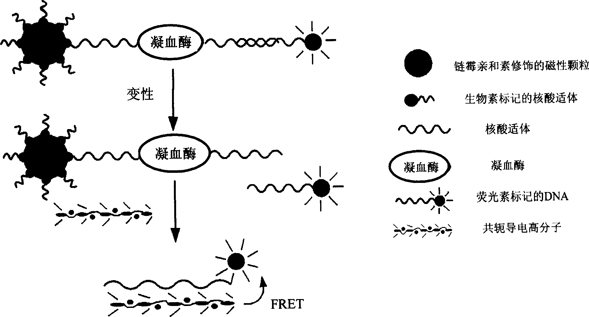
The invention discloses a method which is used for detecting and quantifying target material and based on nucleic acid aptamer and a solid-phase biosensor used for detecting, wherein, the method comprises the following steps: (1) capture probes are connected to the surface of magnetic particle, and the sequence of the capture probes comprises the nucleic acid aptamer sequence of the target material; (2) a sample to be tested is added for reaction; (3) signal probes and bridge molecules are hybridized to obtain double-stranded nucleic acids, and the signal probes are added into the reaction system obtained by step (2) through a double-stranded mode for reaction, the magnetic separation is then carried out to remove free signal probes; (4) the signal probes are separated from the surface of the magnetic particle by a nucleic acid denaturation way, and the fluorescent signals on the signal probes are detected. The method is suitable for detecting wide target materials comprising protein, organic micromolecule, metallic ions, and the like. The method not only has higher sensitivity, but also solves the nonspecific problem and has important meanings in bioinstrumentation, early diagnosis and treatment of disease.

Description

technical field [0001] The invention belongs to the field of biosensors, in particular to a nucleic acid aptamer-based method for detecting and quantifying target substances and a solid-phase biosensor for detection. Background technique [0002] Protein is one of the important components of organisms, and the detection and quantification of some specific proteins is of great significance in disease diagnosis, treatment and new drug development. At present, antibody-based protein detection methods (such as ELISA, Western Blotting) are commonly used in research and medical diagnosis. These traditional methods have the advantages of good specificity and high sensitivity, but often require radioactive labeling, gel electrophoresis and autoradiography. It is not suitable for rapid and high-throughput multiplex detection. The recently developed nucleic acid aptamer (Aptamer) not only has a high specificity similar to antibodies, but also can be screened and can be directly synth...

Claims


Application Information

Patent Timeline
IPC IPC(8): G01N33/551G01N33/53G01N33/68G01N33/573
Inventor 王丽华刘兴奋樊春海
Owner SHANGHAI INST OF APPLIED PHYSICS - CHINESE ACAD OF SCI
Why Eureka
Try Eureka