
Method for separating human spermatogonial stem cells

A technology of spermatogonial stem cells and cells, which is applied in the field of bioengineering, can solve the problems of few isolated cells and low purity, and achieve the effect of high purity and high yield

Inactive Publication Date: 2012-02-08
RENJI HOSPITAL AFFILIATED TO SHANGHAI JIAO TONG UNIV SCHOOL OF MEDICINE
View PDF0 Cites 8 Cited by
  • Summary
  • Abstract
  • Description
  • Claims
  • Application Information

AI Technical Summary

Problems solved by technology

[0005] The technical problem to be solved by the present invention is to provide a method for isolating human spermatogonial stem cells. The method for isolating human spermatogonial stem cells should solve the problem of few isolated cells, Technical problems with low purity

Method used


Image

  • Method for separating human spermatogonial stem cells
  • Method for separating human spermatogonial stem cells
  • Method for separating human spermatogonial stem cells

Examples


Embodiment 1

[0057] A method for separating human spermatogonial stem cells, the steps are as follows:

[0058] 1) Weigh about 1 gram of fresh human testis tissue, place it in 10ml DMEM / F12, and cut it into a volume of about 1×1×1mm 3 Small pieces of tissue were washed 3 times with DMEM / F12 to remove residual blood cells in the tissue.

[0059] 2) Use scissors to cut the testicular tissue to a semi-liquid state, add another 20ml DMEM / F12, and then transfer the tissue to a 150ml container.

[0060] 3) Add digestive enzyme solution I (consisting of a final concentration of 2mg / ml type IV collagenase or type XI collagenase solution and 1μg / μl DNase I) to the shredded tissue in step 2), and place in a 34°C water bath Shake, incubate for 10-15 minutes.

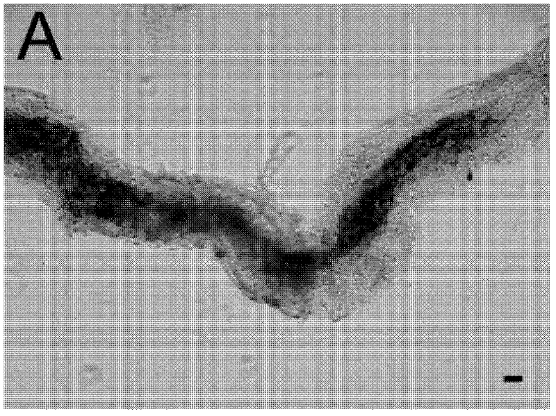
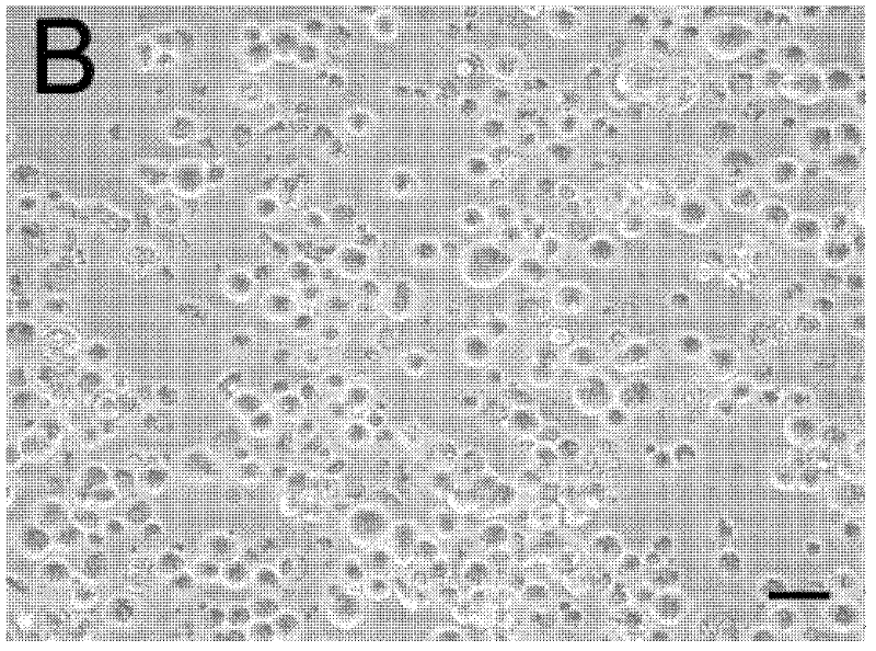
[0061] 4) Microscopic examination to ensure that all tissue pieces have been digested into seminiferous tubules (such as figure 1 shown), transfer the digestion solution containing the seminiferous tubules to another new 50ml centrifuge tube...

Embodiment 2

[0079] Human spermatogonial stem cells were isolated according to Example 1.

[0080] The spermatogonial stem cells were identified by immunocytochemical methods, and the steps were as follows:

[0081] 1) The collected GPR125-positive spermatogonial stem cells or GFRA1-positive spermatogonial stem cells and GPR125- or GFRA1-negative germ cells are subjected to a cell spinner, centrifuged for cell smear, and fixed with 4% paraformaldehyde;

[0082] 2) Wash the cell smear with PBS at room temperature, and block with 200 μl 10% normal goat serum for 30 min;

[0083] 3) Dilute the primary antibody (GPR125, GFRA1, THY1, UCHL1, or ITGA6, or normal rabbit IgG, mouse IgG, negative control with PBS instead of the primary antibody) at 1:200, and incubate at 34°C for 1 hour; after incubation, the cells were incubated with Rinse with PBS;

[0084] 4) Cells were incubated with fluorescein-labeled goat anti-rabbit IgG, or rhodamine-labeled goat anti-rabbit, or rhodamine-labeled goat anti...


PUM


Abstract

The invention belongs to the field of biological engineering and provides a method for separating human spermatogonial stem cells. The method comprises the following steps of: mechanically separating a testicular tissue; processing the testicular tissue into a semi-liquid state; adding digestive enzyme solution I into the liquid testicular tissue in the semi-liquid state; digesting the testiculartissue into a single convoluted tubule; adding digestive enzyme solution II in the convoluted tubule; digesting the convoluted tubule into a single cell; separating reproductive cells from supportingcells in the single cell; separating G protein-coupled receptor 125 (GPR125) positive spermatogonial stem cells from glial cell line-derived neurotrophic factor (GDNF) family receptor alpha 1 (GFRA1)positive spermatogonial stem cells by adopting an immunomagnetic bead cell sorting method; and identifying the separated GPR125 positive spermatogonial stem cells and the GFRA1 positive spermatogonial stem cells by adopting an immune cell chemical method. By the method, a large number of the high-purity human spermatogonial stem cells are obtained, and sufficient cell sources are provided for theresearch of the human spermatogonial stem cells and the application of the human spermatogonial stem cells in regenerative medicine and reproductive medicine.

Description

technical field [0001] The invention relates to the field of bioengineering, in particular to a method for isolating stem cells, in particular to a method for isolating human spermatogonial stem cells. Background technique [0002] Stem cells refer to primitive undifferentiated cells that can both self-renew and differentiate into functional daughter cells. At present, stem cell research has become the frontier of life science research at home and abroad, and is of great significance to cell transplantation, tissue engineering and gene therapy in the treatment of human diseases. Spermatogonial stem cells (SSCs) refer to a subtype of type A spermatogonia. Spermatogonial stem cells are the only adult stem cells that pass genetic material to offspring in male animals and humans. Spermatogonia stem cells are also the basis of spermatogenesis in male animals and humans. The spermatogenesis process in mammals is completed in the seminiferous tubules and includes the complex proc...

Claims


Application Information

Patent Timeline
Patent Type & Authority Applications(China)
IPC IPC(8): C12N5/076
Inventor 何祖平刘阳刘建芳张桢珍李铮杨施郭成志马猛
Owner RENJI HOSPITAL AFFILIATED TO SHANGHAI JIAO TONG UNIV SCHOOL OF MEDICINE
Learn More