Preparation of polylactic-co-glycolic acid (PLGA) nano-fiber scaffold and application of PLGA nano-fiber scaffold to tissue engineering

A nanofiber, tissue engineering technology, applied in the preparation of PLGA nanofiber scaffolds, the application field in tissue engineering, can solve the problems of limiting scaffold/cell in vivo repair effect, difficult to achieve cell differentiation, uneven pore size distribution of cell scaffolds, etc. Achieve uniform distribution, large porosity and sufficient adhesion

Inactive Publication Date: 2013-01-23
JINAN UNIVERSITY
View PDF2 Cites 25 Cited by
  • Summary
  • Abstract
  • Description
  • Claims
  • Application Information

AI Technical Summary

Problems solved by technology

However, at present, tissue engineering scaffolds generally have uneven distribution of cell scaffold pore sizes, and the pore diameter of most cell scaffolds is too small. During the process of culturing cells in vitro, cell

Method used


Image

  • Preparation of polylactic-co-glycolic acid (PLGA) nano-fiber scaffold and application of PLGA nano-fiber scaffold to tissue engineering
  • Preparation of polylactic-co-glycolic acid (PLGA) nano-fiber scaffold and application of PLGA nano-fiber scaffold to tissue engineering
  • Preparation of polylactic-co-glycolic acid (PLGA) nano-fiber scaffold and application of PLGA nano-fiber scaffold to tissue engineering

Examples


Embodiment 1

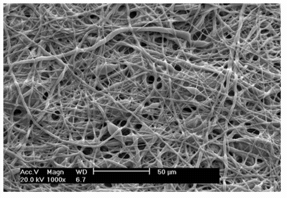
[0037] Prepare a solution of polylactic acid-glycolic acid copolymer (PLGA, LA / GA=75 / 25, Mv=5000, Shandong Institute of Medical Devices) with a mass fraction of 5% in hexafluoroisopropanol as a solvent, and add 5ppm rhodamine B solution, placed in a magnetic stirrer and stirred evenly; using absolute ethanol as a solvent to prepare a polyvinylpyrrolidone (PVP) solution with a mass fraction of 5%, placed in a magnetic stirrer and stirred evenly; draw 0.5mL PLGA solution with a 25mL syringe and 15mL PVP solution, installed in two syringe pumps; cut a piece of tin foil according to the drum of the electrospinning receiving target, and fix it in the drum; open the drum to make it rotate, and adjust the rotation speed of the drum to 20r / min; High-voltage electricity, adjust the voltage intensity to 15KV, adjust the injection speed of PVP solution to 15mL / h, and the injection speed of PLGA solution to 0.5mL / h respectively, so that the two can eject filaments in the electrostatic fiel...

Embodiment 2

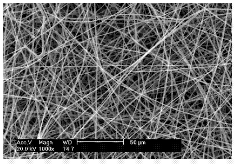
[0040] Prepare a solution of polylactic acid-glycolic acid copolymer (PLGA, LA / GA=75 / 25, Mv=5000, Shandong Institute of Medical Devices) with a mass fraction of 20% in hexafluoroisopropanol as a solvent, and add 8ppm rhodamine B solution, placed in a magnetic stirrer and stirred evenly; using absolute ethanol as a solvent to prepare a polyvinylpyrrolidone (PVP) solution with a mass fraction of 20%, placed in a magnetic stirrer and stirred evenly; use a 25mL syringe to draw 15mL PLGA solution and 0.5 mL of PVP solution, installed in two syringe pumps; cut a piece of tin foil according to the drum of the electrospinning receiving target, and fix it in the drum; open the drum to make it rotate, and adjust the rotation speed of the drum to 100r / min. Turn on the high voltage, adjust the voltage intensity to 20KV, and adjust the injection speeds of the PVP solution and the PLGA solution to 15mL / h and 0.5mL / h respectively, so that the two respectively eject filaments in the electrosta...

Embodiment 3

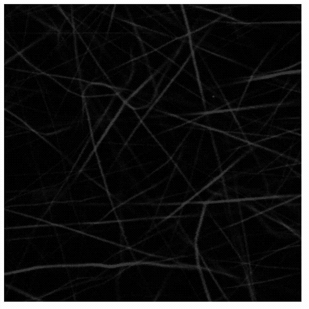
[0043] Prepare a polylactic-co-glycolic acid (PLGA) (PLGA, LA / GA=75 / 25, Mv=5000, Shandong Institute of Medical Devices) solution with a mass fraction of 10% in hexafluoroisopropanol as a solvent, and add 10ppm Rhodamine B solution was placed in a magnetic stirrer and stirred evenly; a solution of polyvinylpyrrolidone (PVP) with a mass fraction of 15% was prepared with absolute ethanol as a solvent, and placed in a magnetic stirrer and stirred evenly; draw 15mL with a 25mL syringe respectively PLGA solution and 3mL PVP solution are installed in two syringe pumps; cut a piece of tin foil according to the drum of the electrospinning receiving target, and fix it in the drum; open the drum to make it rotate, and adjust the rotation speed of the drum to 50r / min ; Turn on the high voltage, adjust the voltage intensity to 25KV, adjust the injection speed of the PVP solution and the PLGA solution to be 10mL / h, so that the two spray filaments in the electrostatic field respectively, and ...


PUM


Abstract

The invention discloses preparation of a polylactic-co-glycolic acid (PLGA) nano-fiber scaffold and application of the PLGA nano-fiber scaffold to tissue engineering, and belongs to the field of the tissue engineering. The method comprises the following steps of: preparing a fiber film containing two materials by taking PLGA and polyvinylpyrrolidone (PVP) as a raw material through electrostatic blended spinning, and dissolving and removing the PVP in an aqueous solution to obtain the PLGA nano-fiber scaffold with certain microstructures. A cell/PLGA nano-fiber scaffold composite matrix is obtained by the following steps of: inoculating seed cells on the PLGA nano-fiber scaffold, culturing in vitro, folding a cell/PLGA nano-fiber scaffold composite, and culturing for 7 to 14 days in a spinner cultivation mode. The composite matrix can be taken as a tissue engineering material with a specific tissue repair function and implanted into an animal model. The preparation process is simple and feasible, is high in repeatability, and can be widely applied to the field of the tissue engineering.

Description

technical field [0001] The invention belongs to the field of tissue engineering and relates to a tissue engineering scaffold, in particular to a preparation method of a PLGA nanofiber scaffold and its application in tissue engineering. Background technique [0002] Tissue engineering is currently a common method for repairing human organs and tissue defects, and its core is to construct a complex composed of cells and scaffolds. Tissue cells are cultured in vitro on scaffolds made of biomaterials that have the same configuration as the tissue or organ to be repaired, and then implanted into the lesion area in the body to allow them to continue to proliferate and differentiate, and finally achieve tissue or organ repair , reconstruction and functional restoration. Therefore, scaffolds prepared from biomaterials play a vital role in tissue engineering. Tissue engineering scaffolds not only provide a three-dimensional space for cell proliferation and differentiation, gas exch...

Claims


Application Information

Patent Timeline
IPC IPC(8): A61L27/18A61L27/16A61L27/38A61L27/60
Inventor 谢德明郭瑞
Owner JINAN UNIVERSITY
Why Eureka
Try Eureka