Antigenic epitope displaying method based on single domain antibody

A single domain antibody, antigen epitope technology, applied in the biological field, can solve the problems of poor epitope peptide immunogenicity, high cost, complex epitope peptide immunogen technology, etc., and achieve the effect of avoiding technical complexity

Active Publication Date: 2013-03-06
BEIJING VICNOVO SCI TECH
View PDF1 Cites 7 Cited by
  • Summary
  • Abstract
  • Description
  • Claims
  • Application Information

AI Technical Summary

Problems solved by technology

[0009] The purpose of the present invention is to establish a new antigenic epitope display method to realize the effective display of antigenic epitopes, to solve th

Method used


Image

  • Antigenic epitope displaying method based on single domain antibody
  • Antigenic epitope displaying method based on single domain antibody
  • Antigenic epitope displaying method based on single domain antibody

Examples


Embodiment 1

[0068] Example 1 Single-domain antibody skeleton display based on camel heavy chain antibody (His) 6 gauge

[0069] Enter the PDB database (http: / / www.pdb.org / pdb / home / home.do), enter "camel antibody" in the search box, and find a camel antibody compounded with lysozyme from the structural data in the search box Structural data (1BZQ) of the object, from which the amino acid sequence of a camelid single domain antibody is shown in SEQ ID NO: 1 (as shown in figure 1 Shown, wherein the double dashed part is the CDR3 region).

[0070] Will figure 1 The amino acid sequence of the CDR3 region shown in is replaced by the "HHHHHH" epitope (His) 6 Epitope, the sequence after replacement is shown in SEQ ID NO: 2 (see attached figure 2 ).

[0071] Then, according to the codon usage bias of Escherichia coli, the sequence shown in the above SEQ ID NO: 2 was converted into its coding gene sequence (http: / / www.bioinformatics.org / sms2 / rev_trans.html), and at the same time, the upstream...

Embodiment 2

[0090] Example 2 Human single domain antibody backbone displaying c-myc epitope and its application

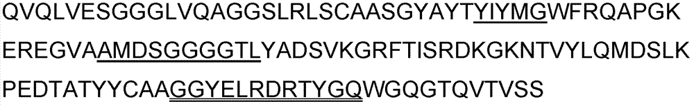
[0091] Obtain a human antibody heavy chain variable region amino acid sequence from the GenBank database and literature (J Exp Med 2011, 208(1): 181-193), as shown in SEQ ID NO: 4, and use this sequence as a single domain antibody base sequence. (if attached Figure 5 Shown, wherein the double underlined part is the framework region 2 (FR2); the single underlined part is the CDR3 region).

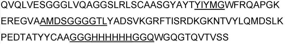
[0092] will attach Figure 5 The 9th, 10th and 12th amino acids in FR2 of the single domain antibody sequence shown in are replaced with glutamic acid (E), arginine (R) and glycine (G) respectively, and at the same time, their The CDR3 region was replaced with the c-myc epitope sequence, namely "EQKLISEEDL", and the c-myc epitope display protein was constructed as shown in SEQ ID NO: 5, (see attached Image 6 shown).

[0093] Then, convert the above sequence into its coding gene sequen...

Embodiment 3

[0113] Example 3 Display of Human Vascular Endothelial Growth Factor Epitope Using the Single-Domain Antibody Skeleton Based on Sharklein Heavy Chain Antibody

[0114] From the literature (Molecular Immunology 2007; 44:656-665), a shark single domain antibody was found, the amino acid sequence of which is shown in SEQ ID NO: 7, (see Figure 9 As shown, the underlined part is the CDR3 region).

[0115] Will Figure 9 The amino acid sequence of the CDR3 region shown in is replaced by human vascular endothelial growth factor (VEGF) antigen epitope "YPDEIEYIFKP" epitope, and the sequence after replacement is shown in SEQ ID NO: 8, (see appendix Figure 10 , where the underlined part is the epitope of human vascular endothelial growth factor).

[0116] Then, according to the codon usage bias of Escherichia coli, the sequence shown in the above SEQ ID NO: X was converted into its coding gene sequence (http: / / www.bioinformatics.org / sms2 / rev_trans.html), and at the same time, the up...


PUM


Abstract

The invention belongs to the technical field of biology and specifically relates to an antigenic epitope displaying method based on a single domain antibody. Specifically, an exogenous antigenic epitope is displayed in a CDR3 region of the single domain antibody, and the single domain antibody skeleton protein which displays the exogenous antigenic epitope on the surface of a molecule can be used for preparing an antibody with a too small epitope, be also used as a substitutive antigen of an exogenous antigen and used for immunizing animals and preparing antibodies aiming at the exogenous antigen. In addition, the antigenic epitope displaying method based on a single domain antibody skeleton can also be used for preparing epitope vaccins.

Description

technical field [0001] The invention belongs to the field of biotechnology, and in particular relates to a method and application of using a single domain antibody to display an exogenous antigen epitope. Background technique [0002] Epitope is a part of protein antigen and is the basis of protein antigenicity, and the specificity of antibody is directed against epitope. Since the epitope is generally only composed of a few to a dozen amino acids, the synthesized individual epitope peptide generally has no or only very weak immunogenicity due to its small molecule. In order to enhance the immunogenicity of antigenic epitope peptides, epitope peptides are generally coupled to carrier proteins such as bovine serum albumin, keyhole limpet hemocyanin, tetanus toxoid, etc., or to lipids with adjuvant activity Molecules are covalently linked to form lipopeptides, and antigenic epitope peptides can also be linked to branched polylysine backbones to form a macromolecule with a uni...

Claims


Application Information

Patent Timeline
IPC IPC(8): C07K19/00C07K1/107C12N15/62C12N15/10C12N15/70
CPCC07K1/107C12N15/1044C07K2317/569C07K19/00C12N15/10C07K16/00C12N15/70C12N15/62
Inventor 高建恩罗时伟钱军
Owner BEIJING VICNOVO SCI TECH
Why Eureka
Try Eureka