Miniature bifocus objective lens probe

A bifocal and microscopic objective lens technology, which is applied in the field of devices in the field of optical technology, can solve the problems of no imaging, no axial scanning ability, and entering the human body, etc., and achieve the effect of small size

Active Publication Date: 2015-05-20
HUAZHONG UNIV OF SCI & TECH
View PDF3 Cites 0 Cited by
  • Summary
  • Abstract
  • Description
  • Claims
  • Application Information

AI Technical Summary

Problems solved by technology

"The disadvantages are as follows: 1. The outer diameter of the design is 7 mm, which cannot enter the human body through the working channel of a conventional endoscope; 2. The probe has no axial scanning ability, and cannot simultaneously analyze the cell morphology of the surface layer of the gastric mucosa and the deep glandular tissue. imaging

Method used


Image

  • Miniature bifocus objective lens probe

Examples


Embodiment Construction

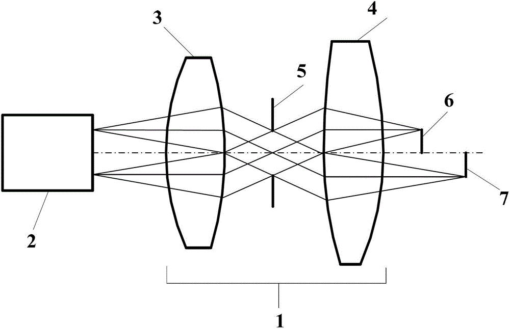
[0016] The invention comprises a miniature bifocal microscope objective lens group 1 and an imaging fiber bundle 2 . The imaging fiber bundle 2 is arranged in front of the miniature bifocal microscope objective lens group 1 . The pupil of the miniature microscopic objective lens group 1 is at infinity, with bifocal imaging capability, the focal lengths are 50 μm and 200 μm, respectively, and can simultaneously image the shallow layer 6 and the deep layer 7 of the sample, and the numerical apertures are 0.5 and 0.4 respectively. The numerical aperture of the miniature microscopic objective lens group in the image space matches the numerical aperture of a single fiber of the imaging fiber bundle.

[0017] The miniature bifocal microscope objective lens group 1 includes a collimating lens unit 3 , a focusing lens 4 and an aperture stop 5 . The collimator lens unit 3 is behind the fiber bundle 2 and before the aperture stop 5; the aperture stop 5 is behind the collimator lens uni...


PUM


Abstract

The invention relates to a miniature bifocus objective lens probe for a confocal endoscopic microscope, belonging to the technical field of optics. The miniature bifocus objective lens probe comprises a miniature bifocus microobjective group and an imaging optic fiber bundle, wherein the imaging optic fiber bundle is arranged in front of the miniature bifocus microobjective group. The pupil of the miniature bifocus microobjective group is at infinity, has a bifocus imaging capability and can be used for simultaneously imaging a superficial layer and a deep layer of a sample. The numerical aperture of the miniature bifocus microobjective group at the optical fiber bundle end is matched with that of single optical fiber of the imaging optical fiber bundle. According to the miniature bifocus objective lens probe, the size is limited within a range that the miniature bifocus objective lens probe can enter the human body through a conventional endoscope working channel, light is focused on two focal planes with different depths, and fluorescent light generated by the focal planes is collected and then returned into a confocal endoscopic imaging system.

Description

technical field [0001] The invention relates to a device in the field of optical technology, in particular to a miniature bifocal objective lens probe suitable for confocal endoscopes. Background technique [0002] In my country, the morbidity and mortality of gastrointestinal tumors rank among the forefront of malignant tumors, and early diagnosis can effectively improve the survival rate. Simultaneously detecting the morphology of mucosal surface cells and deep microvascular structures and obtaining histological imaging results can help in the early diagnosis and classification of gastrointestinal tumors. Confocal endoscopic microscopy is one of the latest endoscopic imaging technologies. It can perform in vivo high-resolution imaging during ordinary endoscopic examination, obtain real-time histological imaging results of the mucosa, and provide doctors with clinical diagnosis and cancer prognosis. provide help. The micro-objective probe is one of the key technologies of...

Claims


Application Information

Patent Timeline
Patent Type & Authority Patents(China)
IPC IPC(8): G02B23/26G02B21/02G02B15/00A61B1/00
Inventor 付玲杨莉张红明刘谦袁菁
Owner HUAZHONG UNIV OF SCI & TECH
Why Eureka
Try Eureka