Anti-malignant lymphoma fusion protein and preparation method thereof

A technology of fusion protein and activation protein, applied in antitumor drugs, peptide/protein components, chemical instruments and methods, etc., can solve problems such as large toxic and side effects, and achieve the effects of high expression, increased productivity, and increased cytotoxicity

Inactive Publication Date: 2013-06-26
ARMY MEDICAL UNIV
View PDF3 Cites 3 Cited by
  • Summary
  • Abstract
  • Description
  • Claims
  • Application Information

AI Technical Summary

Problems solved by technology

So far, the main methods used to treat these diseases are still radiotherapy and chemotherapy, but radiotherapy and chemotherapy kill tumor cells and normal cells at the same time, with extremely toxic side effects

Method used


Image

  • Anti-malignant lymphoma fusion protein and preparation method thereof
  • Anti-malignant lymphoma fusion protein and preparation method thereof
  • Anti-malignant lymphoma fusion protein and preparation method thereof

Examples


Embodiment 1

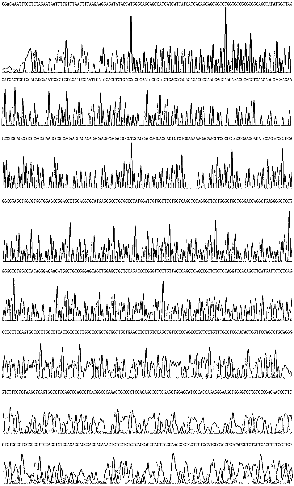
[0033] Example 1 Cloning of Human SARI Gene

[0034] 1.1 Isolation of human peripheral blood mononuclear cells

[0035] (1) Take 5ml of anticoagulated blood and mix it with PBS buffer 1:1.

[0036] (2) Take 10 ml of lymphocyte separation fluid (China TBD Biotechnology Development Center).

[0037] (3) Use a dropper to carefully add the mixture obtained in (1) to the liquid surface of the separation liquid obtained in (2), and let stand at room temperature for 5 minutes.

[0038] (4) Centrifuge horizontally at 2000 rpm for 20 minutes to collect the buffy coat.

[0039] (5) The cells were washed twice with PBS buffer, centrifuged at 2000 rpm for 10 minutes, and the supernatant was discarded. The resulting pellet contained human peripheral blood mononuclear cells.

[0040] 1.2 Extraction of total cellular RNA

[0041] Trizol was used to extract total cellular RNA, and the operation steps were carried out according to the instructions of Trizol reagent.

[0042] (1) In the ...

Embodiment 2

[0061] Example 2 Induced expression and identification of target protein

[0062] 1. Expression of target protein

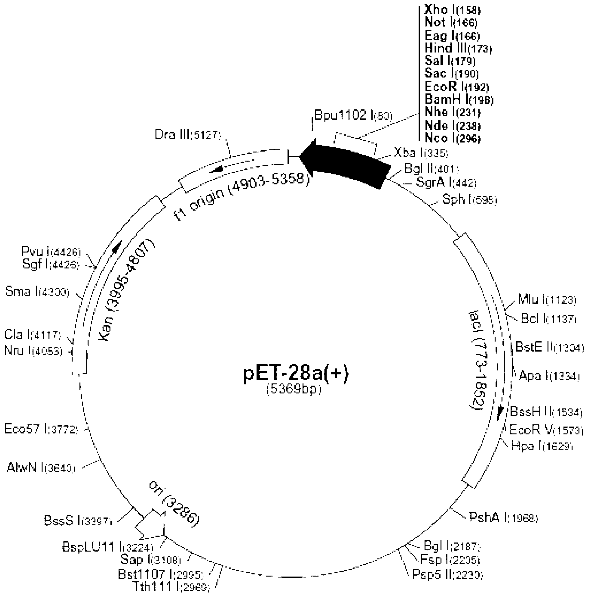
[0063] (1) Pick a single colony containing the recombinant prokaryotic expression plasmid pET-SGRB and the control plasmid pET28a, and inoculate them in 3ml LB liquid medium (containing 50μg / ml Amp), respectively, and culture overnight at 37°C and 220rppm with shaking.

[0064] (2) Inoculate 1% of the bacterial solution cultured by shaking overnight into 5ml LB liquid medium (containing 50μg / ml Amp), shake and culture at 37°C and 250r / min for 3 hours until mid-logarithmic phase [OD(600) ≈0.6].

[0065] (3) Add IPTG to a final concentration of 1 mmol / L, induce expression at 37°C and 220r / min for 4 hours.

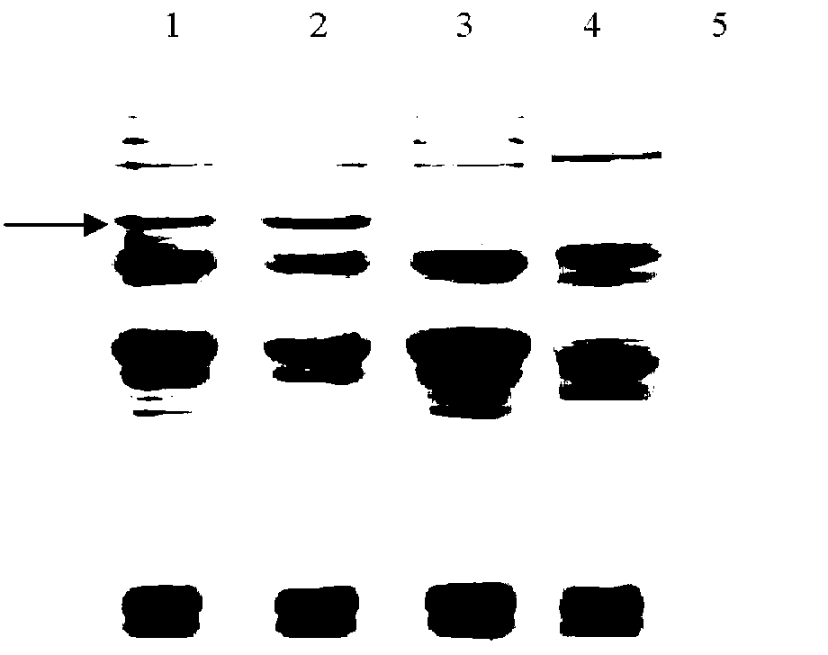
[0066] (4) SDS-PAGE to detect the expression of the target protein.

[0067] 2. Denaturing polyacrylamide gel electrophoresis (SDS-PAGE)

[0068] (1) Take 1ml of the bacterial solution after induction of expression, centrifuge at 10000×g for 1min, and disc...

Embodiment 3

[0097] Example 3 Isolation and purification of target protein

[0098] 1. Mass expression of target protein

[0099] (1) Escherichia coli containing the recombinant prokaryotic expression plasmid pET-SGRB was inoculated in 5 ml of LB culture solution (containing 50 μg / ml Amp), shaken overnight at 37° C. and 220 rpm.

[0100] (2) Inoculate 500ml of LB culture solution containing 100μg / ml ampicillin at 1% of the bacterial solution shaken overnight, shake vigorously at 37°C for 3 hours, until mid-logarithmic phase [D(600)≈0.6].

[0101] (3) Add IPTG to a final concentration of 1 mmol / L.

[0102] (4) Continue shaking at 20°C for 24 hours.

[0103] (5) Bacteria were collected by centrifugation at 4000g for 15 minutes at 4°C.

[0104] 2. Identification of recombinant protein expression

[0105] (1) Add 3ml TE buffer (pH8.0) per gram of bacteria.

[0106] (2) Sonicate the bacteria in an ice bath until the viscous bacterial solution becomes clear.

[0107] (3) Centrifuge at 4°...


PUM


Abstract

The invention provides an anti-malignant lymphoma fusion protein. The anti-malignant lymphoma fusion protein comprises an inhibitor SARI (Suppressor of AP-1 Regulated by Interferon) of a human activator protein-1 and a mutant protein msBAFF of human B cell activating factor BAFF, wherein the msBAFF is a mutant which is formed after amino acids at the 217-224 loci of the human BAFF are replaced by two glycines. The invention further provides a preparation method of the fusion protein. After the recombinant fusion protein of the invention is obtained through a genetic engineering strategy, vitro anti-tumor effect detection is performed and shows that the tumor growth can be effectively inhibited in cells and animals.

Description

technical field [0001] The present invention relates to the fields of biotechnology and biopharmaceuticals. Specifically, the present invention relates to human potent inhibitor SARI (Suppressor of AP-1, Regulated by IFN) protein and B cell activating factor belonging to the TNF family, An anti-malignant lymphoma fusion molecule composed of the mutated protein msBAFF of BAFF), and a preparation method thereof. Background technique [0002] Malignant lymphoma seriously threatens human life and health. So far, the main methods used to treat these diseases are still radiotherapy and chemotherapy, but radiotherapy and chemotherapy kill tumor cells and normal cells at the same time, with extremely toxic side effects. Tumor biotherapy is a promising research field, which plays an important role in overcoming the toxic and side effects of tumor treatment and improving the treatment efficiency. In recent years, the research on AP-1 and BAFF has provided a new way for the realizati...

Claims


Application Information

Patent Timeline
Patent Type & Authority Applications(China)
IPC IPC(8): C07K19/00C12N15/62C12N15/70A61K38/17A61K47/48A61P35/00
Inventor 何凤田张立杨朝辉
Owner ARMY MEDICAL UNIV
Why Eureka
Try Eureka