A Pulmonary Parenchyma Segmentation Method Based on Parabola Modified Convex Hull

A technology of lung parenchyma and parabola, which is applied in the field of medical image processing, can solve problems such as being easily affected by noise, overcompensation, and the effect of repair results, so as to meet the needs of image segmentation and solve the effect that it is difficult to correctly segment

Inactive Publication Date: 2015-09-30
HARBIN ENG UNIV
View PDF3 Cites 3 Cited by
  • Summary
  • Abstract
  • Description
  • Claims
  • Application Information

AI Technical Summary

Problems solved by technology

The existing correction algorithms mainly include: the rolling ball algorithm proposed by Armato S.G., etc., this method uses a ball with a radius R to roll along a certain direction on the edge of the extracted lung area, and judges whether the points in the ball are mutually compatible Eight connected domains are used to judge whether the edge needs to be repaired, but the radius R of the sphere will have a great impact on the repair result. At the same time, the selection of the R value is usually based on experience, which reduces the applicability of the algorithm and often causes overcompensation. Phenomenon
The mathematical morphology method proposed by Hu S. et al. segmented the lung parenchyma. This method has high computational complexity and will affect the accuracy of the edge.
Bollotti R. et al. selected the Glued Elastic Band (GEB) algorithm to repair the lung boundary, imitating the mechanical principle to include the concave part with a smaller bending radius in the required area, while the part with a larger bending radius is still excluded. In addition, this method is computationally complex, and the repair result is also affected by the sampling step size
In addition, Kanazawa K et al. proposed a curvature-based method. According to the large curvature change in the missing part of the lung parenchyma, the curvature threshold is set and the missing part is corrected, but the curvature on the lung boundary is easily affected by noise. impact, and a large amount of calculation
Wang Jing uses the method of endpoint detection, but only for nodules that are not too small or too large (3mm)
Yuan Kehong et al proposed to correct the original lung contour based on the calculation of the local convex hull, and re-include the lung nodules that were originally excluded by mistake, which can effectively correct the initial edge error caused by high-density nodules and blood vessels in the lung, but Due to the need to set multiple parameters, the automatic adaptability to the nodule size is poor, and due to the characteristics of the convex hull algorithm, it is not suitable for the detection of nodules on the inner edge of the lung lobe (there are many natural physiological depressions on the inner edge, such as blood vessels) , heart), it is difficult to obtain a good segmentation effect, and the result of repairing with a straight line cannot completely include the nodules inside the edge

Method used


Image

  • A Pulmonary Parenchyma Segmentation Method Based on Parabola Modified Convex Hull
  • A Pulmonary Parenchyma Segmentation Method Based on Parabola Modified Convex Hull
  • A Pulmonary Parenchyma Segmentation Method Based on Parabola Modified Convex Hull

Examples


Embodiment Construction

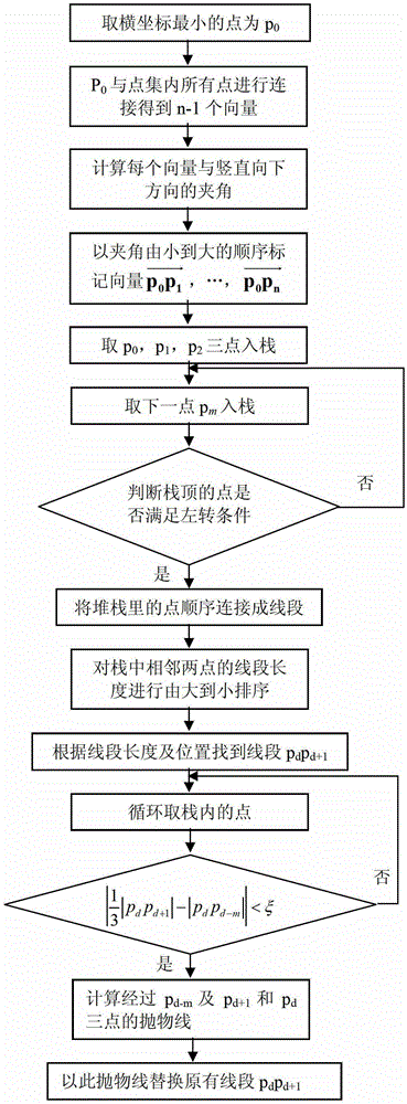
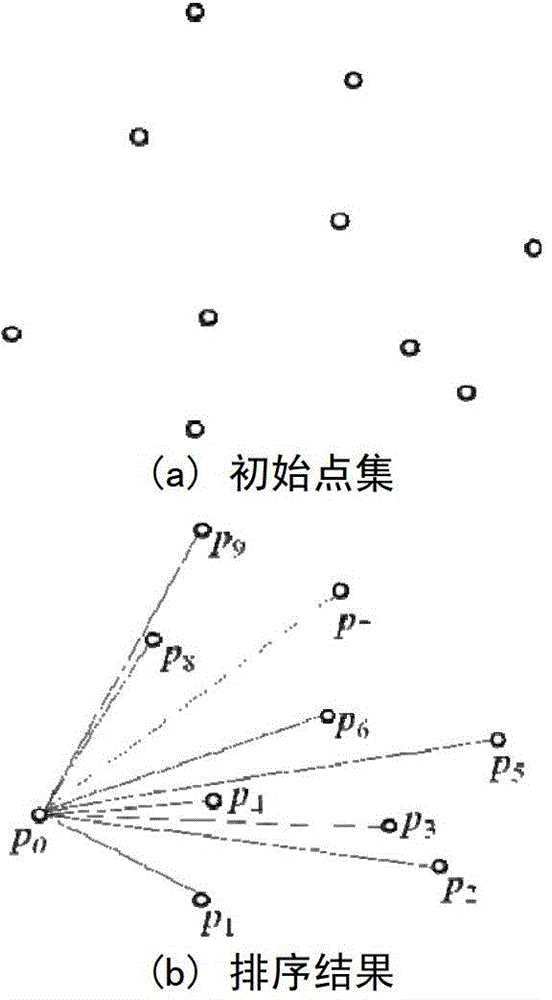
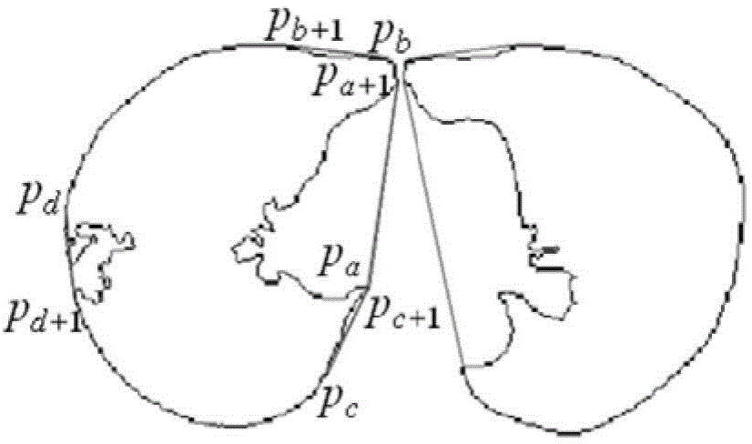
[0028] In this embodiment, a total of 200 clinical chest CT image data conforming to the DICOM3.0 standard from the hospital are used, and all of them have lesions of different degrees. The image size is 440*440, the number of layers is 100-120, and the layer thickness is 2mm. First, the threshold method is used for rough segmentation to obtain a binary image; then, the background is removed by region growth and connected domain judgment to obtain the mask pattern of the lung parenchyma; the contour of the lung parenchyma is obtained by using the edge tracking algorithm; The two-dimensional convex hull algorithm uses a parabola to correct the missing part of the edge; again, the region growing and mathematical morphology operations are used to remove the heart part, and the final mask image is obtained; the mask image and the original image are mathematically operated to obtain a complete of the lung parenchyma.

[0029] 1. Rough extraction of lung parenchyma contour

[0030...


PUM


Abstract

The invention relates to a lung parenchyma segmentation method based on convex hulls correcting by parabolic. The lung parenchyma segmentation method includes extracting lung parenchyma contour images, restoring lung parenchyma external contour and restoring lung parenchyma internal edges sequentially. The lung parenchyma segmentation method is characterized in that lung parenchyma external contour restoration is realized through the steps of 1, ranking points in lung parenchyma contour image edge contour; 2, establishing edge stacks of points in step one, and obtaining an edge point set P= {p0, p1, ... pn} (n> / =3); 3, connecting two adjacent points in the point set P to be segments, ranking the segments in descending order of length, and finding segments pdpd +1 of pulmonary nodules corresponding to missing parts of lobe contour according to segment length and position; 4, utilizing the parabolic to correct the segments pdpd +1, and obtaining lung parenchyma edge contour.

Description

technical field [0001] The invention relates to the field of medical image processing, in particular to a lung parenchyma segmentation method based on a parabola modified convex hull. Background technique [0002] With the development of computer tomography (CT) technology and computer image processing technology, the computer-aided diagnosis system (Computer-Aided Detection) provides a new means for accurate, fast and automatic analysis and processing of CT image data. Help to improve the sensitivity and specificity of doctors' diagnosis. In the study of computer-aided diagnosis of lung diseases, the segmentation of lung parenchyma can eliminate the influence of irrelevant factors in CT images (such as chest, heart, examination bed, etc.), reduce unnecessary calculations, and the accuracy of segmentation is the most important factor. Quantitative analysis of lung disease, detection of pulmonary nodules and key link in lung function assessment. [0003] In CT images, since...

Claims


Application Information

Patent Timeline
Patent Type & Authority Patents(China)
IPC IPC(8): G06T7/00
Inventor 梁洪李金郑冰丛望王磊王可新鲍佩华
Owner HARBIN ENG UNIV
Why Eureka
Try Eureka