Fluorescence microscopy method for rapidly and efficiently observing camellia plant pollen tube

A technology of plant pollen and Camellia, which is applied in the field of fluorescence microscopy, can solve the problems of difficulty in determining the weight of the tablet, affecting the observation effect, and overlapping tissues, so as to shorten the transparent time, improve the experimental efficiency, and avoid the effect of material shrinkage

Active Publication Date: 2013-09-25
CENTRAL SOUTH UNIVERSITY OF FORESTRY AND TECHNOLOGY
View PDF2 Cites 13 Cited by
  • Summary
  • Abstract
  • Description
  • Claims
  • Application Information

AI Technical Summary

Problems solved by technology

In addition, it is difficult to determine the severity of the tableting. If the tableting is too light, the tissues will overlap, which will affect the observation effect; if the tableting is too heavy, the material will be broken, and the grow

Method used


Image

  • Fluorescence microscopy method for rapidly and efficiently observing camellia plant pollen tube
  • Fluorescence microscopy method for rapidly and efficiently observing camellia plant pollen tube
  • Fluorescence microscopy method for rapidly and efficiently observing camellia plant pollen tube

Examples


Embodiment 1

[0046] Embodiment 1 material anatomical separation

[0047] The anatomical separation process of the pistil of Camellia oleifera 'Huasuo' is divided into the anatomical separation of the ovary and the anatomical separation of the style, and the corresponding anatomical separation methods are as follows:

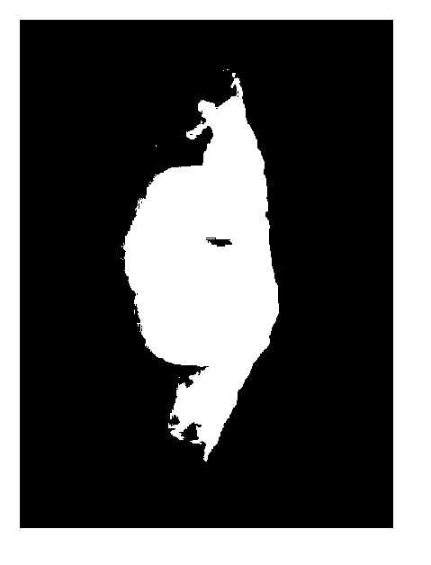
[0048] Anatomical isolation of Camellia oleifera 'Huashuo' ovary: unpollinated or pollinated pistils of Camellia oleifera were removed from the tree, fixed with Carnot's fixative and stored in 70% alcohol. When dissecting, take out the pistil and place it in a petri dish filled with 70% alcohol, and place it in the field of view of the dissecting microscope. Under the dissecting microscope, the style and fruit stalk of Camellia oleifera are excised, the ovary wall is scraped off, and the ovules are exposed in each ventricle (such as Picture 1-1 , Figure 1-2 shown), cut each ventricle from the central axis, and then continue to divide according to the orientation of the ovul...

Embodiment 2

[0053] In a weighing bottle or a penicillin bottle, the separated material to be observed (one operating unit) is rehydrated with various levels of alcohol (50% alcohol → 30% alcohol → distilled water), and in a NaClO solution with an available chlorine content of 9000mg / L Soak for 2 hours, wash with distilled water for 3 times, soak in NaOH solution with a concentration of 8 mol / L for 2 hours, wash with distilled water for 3 times and wash with running water for 30 minutes, in a water-soluble aniline blue dye solution with a mass concentration of 0.2% (with a pH of 8 Prepared with phosphate buffer solution) stained for 6 hours, prepared for observation.

[0054] The specific operation method of "double-slide bonding two-sided observation method" is (such as Figure 3-2 Shown): Take a glass slide A with a thickness of 1.0mm, place the material to be observed after being dyed with water-soluble aniline blue dye on it, drip water-soluble aniline blue dye around the material, and...

Embodiment 3

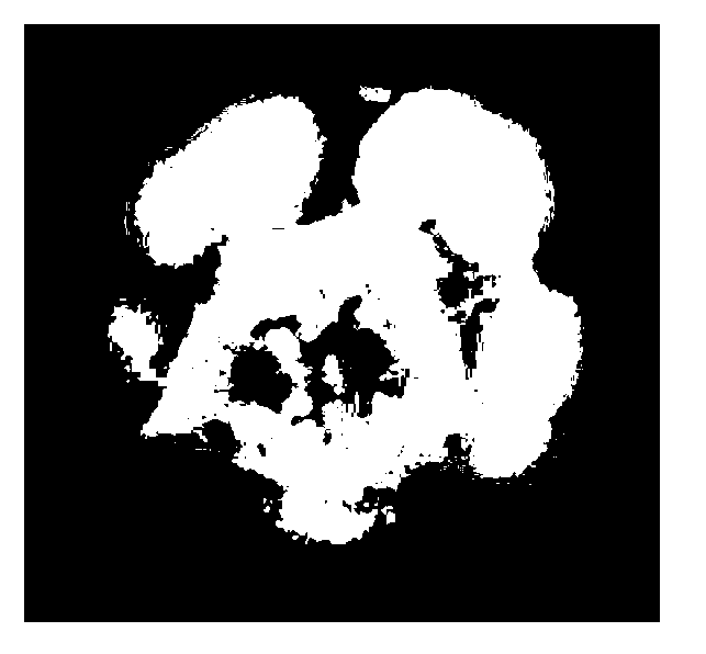
[0056] The ovule material (not pollinated) of camellia oleifera 'Huashuo' separated according to the method in Example 1 was made transparent by NaClO solution, softened by NaOH solution, stained with aniline blue, and prepared by "double-slide bonding and two-sided observation method", and the effect was observed by fluorescence microscope Such as Pic 4-1 shown.

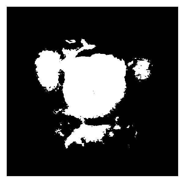
[0057] The ovule material (not pollinated) of Camellia oleifera 'Huasuo' separated according to the method in Example 1 was softened by NaOH solution and stained with aniline blue in the traditional observation method, and then prepared by the "double-slide bonding two-sided observation method" (except that there is no NaClO Except the step that the solution is transparent, other steps are the same as the method described in Example 2), and the observation effect by fluorescence microscope is as follows: Figure 4-2 shown.

[0058] Compared Pic 4-1 and Figure 4-2 It can be seen that using the method of the pr...


PUM


Abstract

The invention relates to a fluorescence microscopy method for rapidly and efficiently observing a camellia plant pollen tube. The method comprises the following steps: (1) dissecting and separating a pistil material of a camellia plant to be observed into an operation unit; and (2) sequentially soaking the separated material in NaClO solution and NaOH solution until the material is transparent and gets softened, dyeing through a water-soluble aniline blue dye solution, flaking by employing a double-glass slide attachment both-sided observation method, and finally performing pollen tube fluorescence microscopy. The method is easy, convenient and feasible to operate, the experiment efficiency is greatly improved, and the observation effect is optimized.

Description

technical field [0001] The invention relates to the technical field of plant microscopy, in particular to a fluorescence microscopy method for quickly and efficiently observing pollen tubes of Camellia plants. Background technique [0002] The pollen tube fluorescence microscopic observation method uses low concentration of water-soluble aniline blue dye (aniline blue dye contains fluorescent pigment) to impregnate plant materials under alkaline conditions, so that the fluorescent dye adsorbed on plant cells is excited by ultraviolet light from a fluorescence microscope. When the excitation light is absorbed by the material, fluorescence is generated and released, which visually shows the growth track of the pollen tube. [0003] In the traditional pollen tube fluorescence microscopy technique, the plant material (such as style or ovary) is first softened, and then dip-dyeed with aniline blue dye, and the fluorescent observation is carried out after the combination of "cover...

Claims


Application Information

Patent Timeline
IPC IPC(8): G01N21/64
Inventor 袁德义高超杨亚邹锋
Owner CENTRAL SOUTH UNIVERSITY OF FORESTRY AND TECHNOLOGY
Why Eureka
Try Eureka