


Quick Research
Generate reliable direction feasibility study reports for your R&D in just a few steps.

Technical Q&A
Discover and master advanced knowledge NOW. Basics, ideas, possibilities, all at once.

Find Solutions
As an expert in R&D theories, this can generate solutions to your technical problems instantly.

Evaluate Feasibility
Analyze your overall solution with one click, know your potential R&D risks in advance.

Monitor Landscape
Get weekly tech updates, stay abreast of the latest tech innovations and key insights.
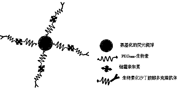
Method for marking salbutamol polyclonal antibody by fluorescent microsphere
A technology of polyclonal antibodies and fluorescent microspheres, which is applied in the direction of measuring devices, instruments, scientific instruments, etc., can solve the problems of low antibody labeling efficiency and label dispersion, and achieve good colloidal stability, good dispersibility and solubility, and binding strong effect
- Summary
- Abstract
- Description
- Claims
- Application Information
AI Technical Summary
Problems solved by technology
Method used
Image

Examples
Embodiment 1
[0020] Example 1: Preparation method of novel conjugates of fluorescent microsphere-labeled salbutamol polyclonal antibody in the present invention
[0021] 1) Preparation method of amino-modified fluorescent microspheres
[0022] Wash 10 mg of fluorescent microspheres and ultrasonically dissolve them in 1 mL of distilled water, add 20 μL of glacial acetic acid and 20 μL of (3-trimethoxysilylpropyl)-diethylethylenediamine for ultrasonic dissolution, and stir magnetically for 3 to 4 h, washed with 10 mM MES buffer (pH 5.5) and resuspended.
[0023] 2) Preparation method of active esterified biotin
[0024] Accurately weigh 4.5 mg biotin-PEG 5000 The complex was dissolved in 2 mL of DMF solution, and after adding 20.5 mg of dicyclohexylcarboimide and 10.5 mg of N-hydroxysuccinimide, the reaction was carried out under magnetic stirring at room temperature for 24 h, and then centrifuged at 4000 rpm for 5 min, and the precipitate was discarded. Actively esterified biotin was o...
Embodiment 2
[0028] Example 2: Application of a novel conjugate of fluorescent microsphere-labeled albuterol polyclonal antibody on fluorescent microsphere test strips
[0029] 1) Preparation of fluorescent microsphere immunochromatographic test strips
[0030]Preparation of fluorescent microsphere pads: After reconjugating the conjugate prepared in Example 1 with 0.01 M PNPB (which contains 5% sucrose and 0.05% Tween-20) to the initial volume, use BIODOT Dispensing System, according to 4 μL The amount of / cm sprayed onto the glass fiber membrane, vacuum dried at 25 ℃ for 1-2 hours, and placed in a dry environment for later use.
[0031] The albuterol-BSA conjugate and the donkey anti-mouse secondary antibody were coated on the NC membrane: adjust the coating concentration to 0.4 mg / mL and 0.4 mg / mL with 0.01 M PBS buffer respectively, and spray the membrane volume to 0.74 μL / cm, the detection line is coated with albuterol-BSA conjugate, the quality control line is coated with donkey a...
Embodiment 3
[0040] Example 3: Detection of salbutamol residues in pig liver on fluorescent microsphere test strips prepared by a novel conjugate of salbutamol polyclonal antibody labeled with fluorescent microspheres
[0041] The sample pretreatment method and test strip production are the same as in Example 2.
[0042] The qualitative and quantitative detection methods are the same as in Example 2, and a standard curve is drawn by adding a series of concentrations (0.5, 1, 2, 3, 4, 5, 6, 8, 10, 15 ng / mL) and corresponding fluorescence intensity values. Draw 100 μL of the extract and add it to the sample well. After 10 minutes, use a reader to detect. According to the data output of the test sample, the output values are 2.90. Check the standard curve to find that the residual amount of salbutamol in the test sample is 0.
PUM
| Property | Measurement | Unit |
|---|---|---|
| Particle size | aaaaa | aaaaa |
Abstract
Description
Claims
Application Information
 - R&D Engineer
- R&D Manager
- IP Professional
- Industry Leading Data Capabilities
- Powerful AI technology
- Patent DNA Extraction
Browse by: Latest US Patents, China's latest patents, Technical Efficacy Thesaurus, Application Domain, Technology Topic, Popular Technical Reports.
© 2024 PatSnap. All rights reserved.Legal|Privacy policy|Modern Slavery Act Transparency Statement|Sitemap|About US| Contact US: help@patsnap.com