AFM probe for single molecular force spectrum analysis and substrate functional modification method
A technology of single-molecule force spectroscopy and modification methods, which is applied in scanning probe technology, scanning probe microscopy, measuring devices, etc., and can solve problems such as complicated steps and increased risk of gold surface functionalization failure
- Summary
- Abstract
- Description
- Claims
- Application Information
AI Technical Summary
Problems solved by technology
Method used
Image

Examples
Embodiment 1
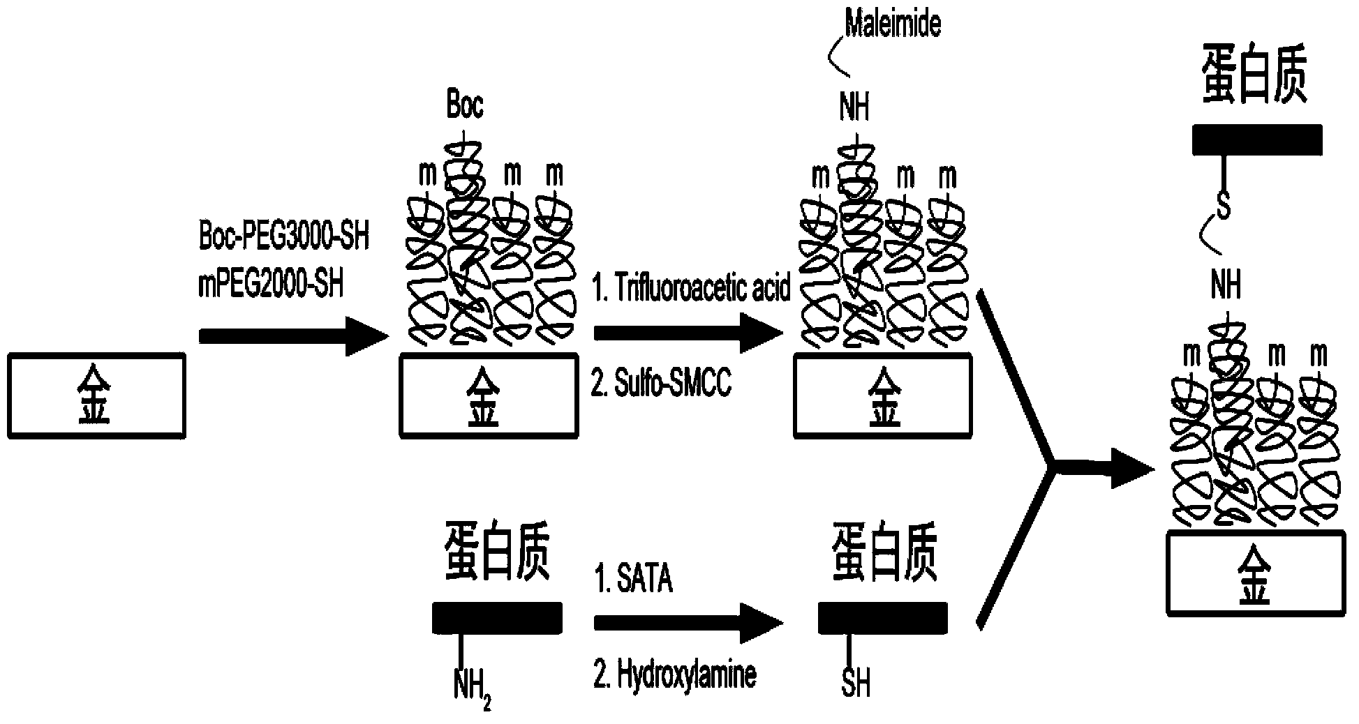
[0025] Functional modification of gold probes: wash the gold-plated atomic force microscope probes (gold probes) in chloroform, dry under nitrogen, wash with ultraviolet and ozone for 10 min, then wash with ultrapure water, wash with chloroform, and dry under nitrogen; The clean gold probe and gold substrate were put into 1 mM Boc-PEG-3000-SH and mPEG-2000-SH mixture (molar ratio 0.5:99.5) in chloroform for 12h, and then added to trifluoroacetic acid React in medium for 5 minutes, wash with ultrapure water, dissolve the cross-linking agent Sulfo-SMCC in ultrapure water, the concentration is 5mg / mL, dilute to 0.5mg / mL with PBS, take 100μL to react with the gold probe for 30min; dissolve SATA to two In methylformamide, the concentration is 1mg / mL, take 1.5μL SATA solution and add dropwise to 100μL 1mg / mL monoclonal anti-goat IgG, react for 2h, then take 3μL deacetylation buffer (0.5M hydroxylamine, 25mM EDTA, PBS buffer system, pH 7.2-7.5) was added dropwise to 30 μL monoclonal ...
Embodiment 2
[0029] Functional modification of gold probes: wash the gold-plated atomic force microscope probes (gold probes) in chloroform, dry under nitrogen, wash with ultraviolet and ozone for 10 min, then wash with ultrapure water, wash with chloroform, and dry under nitrogen; The clean gold probe and gold substrate were put into 1 mM Boc-PEG-3000-SH and mPEG-2000-SH mixture (molar ratio 1:99) in chloroform for 12h, and then added to trifluoroacetic acid React in medium for 5 minutes, wash with ultrapure water, dissolve the cross-linking agent Sulfo-SMCC in ultrapure water, the concentration is 5mg / mL, dilute to 0.5mg / mL with PBS, take 100μL to react with the gold probe for 30min; dissolve SATA to two In methylformamide, the concentration is 1mg / mL, take 1.5μL SATA solution and add dropwise to 100μL 1mg / mL monoclonal anti-goat IgG, react for 2h, then take 3μL deacetylation buffer (0.5M hydroxylamine, 25mM EDTA, PBS buffer system, pH 7.2-7.5) was added dropwise to 30 μL monoclonal anti...
Embodiment 3
[0033] Functional modification of gold probes: wash the gold-plated atomic force microscope probes (gold probes) in chloroform, dry under nitrogen, wash with ultraviolet and ozone for 10 min, then wash with ultrapure water, wash with chloroform, and dry under nitrogen; Clean gold probes and gold substrates were put into 1 mM Boc-PEG-3000-SH and mPEG-2000-SH mixture (molar ratio 5:95) in chloroform for 12 hours, and then added to trifluoroacetic acid React in medium for 5 minutes, wash with ultrapure water, dissolve the cross-linking agent Sulfo-SMCC in ultrapure water, the concentration is 5mg / mL, dilute to 0.5mg / mL with PBS, take 100μL to react with the gold probe for 30min; dissolve SATA to two In methylformamide, the concentration is 1mg / mL, take 1.5μL SATA solution and add dropwise to 100μL 1mg / mL monoclonal anti-goat IgG, react for 2h, then take 3μL deacetylation buffer (0.5M hydroxylamine, 25mM EDTA, PBS buffer system, pH 7.2-7.5) was added dropwise to 30 μL monoclonal a...
PUM
 Abstract
Description
Claims
Application Information
 - R&D
- Intellectual Property
- Life Sciences
- Materials
- Tech Scout
- Unparalleled Data Quality
- Higher Quality Content
- 60% Fewer Hallucinations
Browse by: Latest US Patents, China's latest patents, Technical Efficacy Thesaurus, Application Domain, Technology Topic, Popular Technical Reports.
© 2025 PatSnap. All rights reserved.Legal|Privacy policy|Modern Slavery Act Transparency Statement|Sitemap|About US| Contact US: help@patsnap.com