
Fluorescent endoscope imaging system and method

An imaging system and fluorescence image technology, applied in endoscopy, medical science, diagnosis, etc., can solve the problems of prolonging the exposure time of CCD cameras, large loss of optical signals, and loss of optical signals, etc., to reduce the probability of motion artifacts , reduce the loss of optical signal, shorten the effect of exposure time

Inactive Publication Date: 2014-03-26
GENERAL HOSPITAL OF CHINESE PEOPLES ARMED POLICEFORCES
View PDF9 Cites 26 Cited by
  • Summary
  • Abstract
  • Description
  • Claims
  • Application Information

AI Technical Summary

Problems solved by technology

Since AFI does not use a specific targeting contrast agent, the false positive rate is high, especially for distinguishing inflammation from tumor.
At the same time, because the autofluorescence signal intensity is very weak, it is easily affected by the aliasing of excitation light.
[0012] (2) Shared optical fiber channel fluorescence imaging equipment: This imaging equipment has a simple composition, and the irradiation part of the excitation light coincides with the image acquisition part of the image transmission fiber; The exposure time of the CCD camera needs to be extended, and the probability of artifacts will increase after being affected by gastrointestinal peristalsis, which cannot fully meet the needs of digestive endoscopy imaging
[0013] (3) Parallel dual-fiber channel fluorescence imaging equipment: the image transmission fiber cannot collect all the irradiated parts of the excitation fiber, and at the same time, the image range collected by the image transmission fiber cannot be fully irradiated by the excitation light transmitted by the excitation fiber, resulting in the distortion of the optical signal. loss
[0014] (4) Multispectral imaging cannot be realized
For example, if the excitation light is too concentrated on the area where a certain normal tissue is located, the trace fluorescent probe in the normal tissue will also emit strong fluorescence similar to the diseased tissue, resulting in a false positive
Secondly, because the attenuation rate of the existing commercial optical filters to the light in the stop band is about 99.9%, and the light intensity of the excitation light itself is about 1000 times stronger than that of the fluorescence, the fluorescence filter cannot fully filter the surface of the probe. Scattered excitation light, the excitation light signal still accounts for a large part of the fluorescence image, reducing the signal-to-noise ratio of the fluorescence image, reducing the contrast between diseased tissue and normal tissue on the fluorescence image, resulting in false negatives
[0016] The filters of the three existing fluorescence imaging devices are cured and cannot be easily replaced
Different fluorescent probes require excitation light in different spectral bands, and the emitted fluorescence is also distributed in different spectral bands. Therefore, specific excitation light sources and filter devices are required for certain fluorescent probes. Currently, commonly used fluorescent endoscopes only Excitation imaging of a specific fluorescent probe

Method used


Image

  • Fluorescent endoscope imaging system and method
  • Fluorescent endoscope imaging system and method
  • Fluorescent endoscope imaging system and method

Examples


Embodiment 1

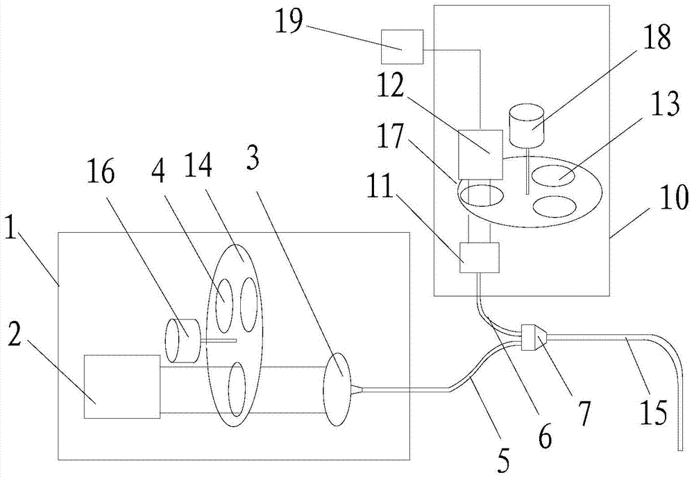
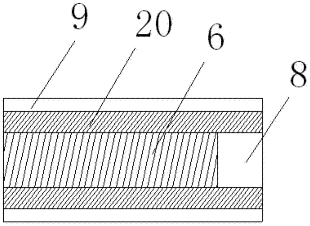
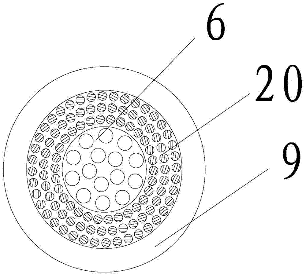
[0051] Refer to attached Figure 1-Figure 3 , the specific embodiments of the present invention will be described below. in, figure 1 Schematically shows the basic structural diagram of the fluorescent endoscopic imaging system of the present invention, and the specific parts are described as follows:

[0052] Light source department

[0053] The light source part is located in the first dark box 1, which generates excitation light of a specific spectrum through the scheme of spectral filtering of the broad-spectrum light source, so as to achieve the purpose of being able to cooperate with various fluorescent probes. It contains a broadband light source 2, a first filter switcher (including a first filter wheel 14, a first wheel controller 16 and an excitation filter mounted on the first filter wheel 14 4) and optical collimation coupler 3. The wide-spectrum light source 2 can generate white light with uniform light intensity distribution in the wavelength range from visib...

Embodiment 2

[0076] This embodiment provides a method for rapid multispectral imaging of two or more fluorescent probes used in the aforementioned fluorescent endoscopic imaging system, so as to use two kinds of IntegriSense645 and RediJect2-DG-750 for the rabbit intestinal cancer model. Taking multispectral imaging with fluorescent probes as an example, the following steps are included:

[0077] S10. According to the spectroscopic characteristics of the two fluorescent probes IntegriSense645 and RediJect2-DG-750 used, select a suitable combination of two sets of excitation light filters 645nm and 750nm and fluorescence filters 720nm and 820nm.

[0078] S20. Align the end of the composite optical fiber bundle 15 with the detection target, turn on the wide-spectrum light source 2, switch the first filter switcher to allow the neutral attenuation sheet to enter the optical path, irradiate the detection object with white light, and switch the second filter switch Connect the port to the optic...


PUM


Abstract

The invention discloses a fluorescent endoscope imaging system and method, which can be used together with most of the conventional fluorescent probes to accurately excite and develop the distribution of fluorescent probes of various disease targeting tissues in real time. The fluorescent endoscope imaging system is provided with a light source part, an optical signal conduction and acquisition part, an image acquisition part and an image denoising and displaying part, wherein the light source part can generate white light and multispectral excitation light matched with the absorption spectrum of the fluorescent probes; the optical signal conduction and acquisition part can be used for conducting the excitation light or the white light onto cavity tissues in a human body, uniformly irradiating the excitation light or the white light onto a detection target, collecting white light and excitation light diffused by a cavity surface in the imaged human body and fluorescent signals emitted under stimulation and conducting an optical signal to the outside of the body; the image acquisition part can be used for acquiring an original fluorescent image, an excitation light image and a white light image in sequence; the image denoising and displaying part is used for removing noise produced by spectrum aliasing in the fluorescent image and displaying a denoised fluorescent image and white light image.

Description

technical field [0001] The invention relates to imaging technology, in particular to a fluorescent endoscopic imaging system and method. Background technique [0002] my country is a country with a high incidence of gastroesophageal tumors. The incidence and death rate of gastric cancer ranks second in my country's large and medium-sized cities, and ranks first in rural areas. Esophageal cancer is also one of the most common tumors in my country. Fluorescent endoscopic imaging technology has a good prospect in the early detection of gastroesophageal tumors. [0003] Fluorescence endoscopic imaging systems in the prior art include the following types: [0004] (1) Autofluorescence imaging equipment [0005] Autofluorescence imaging (AFI) equipment uses xenon light to pass through a blue-green rotary filter to form exciting blue light (wavelength 390-470nm) and green light (wavelength 540-560nm) to directly irradiate the gastrointestinal mucosa, except for the reflected blue...

Claims


Application Information

Patent Timeline
IPC IPC(8): A61B1/05
Inventor 刘海峰何耘屈亚威白净刘飞陆小健徐明泉
Owner GENERAL HOSPITAL OF CHINESE PEOPLES ARMED POLICEFORCES
Learn More