Eureka AIR delivers breakthrough ideas for toughest innovation challenges, trusted by R&D personnel around the world.

Method for improving various bionic functions on surface of cardiovascular implant material

An implant material and surface improvement technology, which is applied in artificial cell constructs, animal cells, medical science, etc., can solve the problems that the shape and function have not been fully demonstrated, the surface blood compatibility is not good, and the anticoagulation effect is not good. Achieve the effect of improving bionic degree, remarkable effect and simple preparation process

Inactive Publication Date: 2014-12-24
SOUTHWEST JIAOTONG UNIV
View PDF8 Cites 2 Cited by
  • Summary
  • Abstract
  • Description
  • Claims
  • Application Information

AI Technical Summary

Problems solved by technology

[0007] The preparation technology of HA micropatterns on the surface of biomaterials is a relatively mature existing technology, which can achieve the purpose of regulating cell morphology, behavior and function by preparing HA micropatterns of different sizes on the surface of materials, but the prepared surface blood phase Poor compatibility, its anticoagulant effect is not even as good as the original material surface; one or two components in the endothelial extracellular matrix are fixed on the surface of biomaterials to achieve the dual effects of anticoagulant and endothelial promotion. This is a mature existing technology, but the disadvantage is that the constructed surface only focuses on the biomimetic of one or two functions, rather than the biomimicry of the complete function of the inner wall of the blood vessel; the relatively complete endothelial extracellular matrix obtained by deendothelial cell technology on the surface of in vitro materials is currently also It has been reported, but the function of immune rejection and inhibition of inflammatory response of this surface has not been described. In addition, the culture of endothelial cells during the surface preparation process has not been carried out under biomimetic conditions. Its morphology, function and secreted extracellular matrix Whether it has complete functions has not been fully demonstrated
However, there is no report on obtaining this multifunctional biomimetic endothelial extracellular matrix by using striped HA micropatterns on the surface of cardiovascular materials to regulate endothelial cell fusion in vitro.

Method used


Image

  • Method for improving various bionic functions on surface of cardiovascular implant material
  • Method for improving various bionic functions on surface of cardiovascular implant material
  • Method for improving various bionic functions on surface of cardiovascular implant material

Examples


Embodiment 1

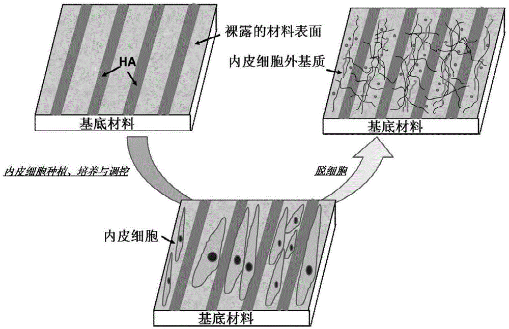
[0027] see figure 1 , the first embodiment of the present invention is a method for obtaining multifunctional vascular endothelial extracellular matrix on a stainless steel surface, the steps of which are:

[0028] A. Prepare a molecular weight of 5×10 on the surface of polished stainless steel 5 DA's hyaluronic acid (HA) striped micropattern, the strip widths of HA and bare material are 35 μm and 15 μm, respectively, for use;

[0029] B. Preparation of vascular endothelial extracellular matrix on the surface of stainless steel: the vascular endothelial cells of the third generation were divided into 5×10 4 The density of each / ml is planted on the surface of the micropattern described in step A, at 37 ° C, 5% CO 2 The concentration of the standard culture conditions was cultivated for 2 days. This step obtained vascular endothelial cells with elongated shape and orderly arrangement after being regulated by HA micropatterns; the used cell culture medium was sucked off, and th...

Embodiment 2

[0032] A method for obtaining multifunctional vascular endothelial extracellular matrix on a titanium surface, the steps of which are:

[0033] A. Prepare a molecular weight of 1×10 on the polished titanium surface 6 DA's hyaluronic acid (HA) striped micropattern, the strip widths of HA and bare material are 25 μm and 25 μm, respectively, for use;

[0034] B. Preparation of vascular endothelial extracellular matrix on titanium surface: the vascular endothelial cells of the second generation were mixed with 1×10 5 The density of each / ml is planted on the surface of the micropattern described in step A, at 37 ° C, 5% CO 2 Under the standard culture conditions of the concentration of 1 day, this step can obtain vascular endothelial cells with elongated shape and orderly arrangement after being regulated by the HA micropattern; aspirate the used cell culture medium, and use 37°C for the sample of cultured cells After washing with PBS 4 times, add decellularization solution, 37°C...

Embodiment 3

[0037] A method for obtaining a multifunctional vascular endothelial extracellular matrix on the surface of a polyurethane material, the steps of which are:

[0038] A. Prepare a molecular weight of 3×10 on the surface of the polyurethane material 6 DA's hyaluronic acid (HA) striped micropattern, the strip widths of HA and bare material are 30 μm and 20 μm, respectively, ready for use;

[0039] B. Preparation of vascular endothelial extracellular matrix on the surface of polyurethane material: the vascular endothelial cells of the third generation were divided into 8×10 4 The density of each / ml is planted on the surface of the micropattern described in step A, at 37 ° C, 5% CO 2 Cultivate for 3 days under the standard culture conditions of HA concentration. This step will obtain vascular endothelial cells with elongated shape and orderly arrangement after being regulated by HA micropatterns; suck out the used cell culture medium, and use 37°C for the samples of cultured cells...


PUM

PropertyMeasurementUnit
widthaaaaaaaaaa

Abstract

The invention discloses a method for improving various bionic functions on the surface of a cardiovascular implant material. The method comprises the following steps: first, preparing an HA (Hyaluronic Acid) stripe-like micrograph with high molecular weight, cultivating vascular endothelial cells on the surface, and regulating in vitro construction of extracellular matrixes of vascular endothelial cells through the HA stripe-like micrograph; and then, peeling the endothelial cells on the surface of the material by a decellularizing technology so as to prepare a multifunctional biological modified surface with anticoagulation / endothelialization promotion / smooth muscle cell adhesion inhibition effects and macrophage adhesion proliferation / inhibition and spreading on the surface of the cardiovascular implant material. The method disclosed by the invention constructs the multifunctional bionic endothelial cell extracellular matrix modified layer which is of anticoagulation and reendothelialization, inhibits adhesion and proliferation of smooth muscle cells, promotes phenotype shrink and inhibits adhesion and spreading of macrophage. The blood compatibility of the material, the structural and functional repair capacity of endothelial injury are remarkably improved.

Description

technical field [0001] The invention belongs to the intersection field of biological material surface engineering and biological imitation manufacturing engineering. Background technique [0002] The surface modification technology of cardiovascular implant materials has gradually matured and is widely used in clinical practice. However, the surface efficacy of materials currently used in clinical practice is far from reaching the expected goal. This is mainly due to the fact that no matter how good the surface modification is, its bionic function cannot match the sound biological function of the blood contact interface in the body's own cardiovascular system. [0003] Through the biomimicry of the surface of the material, endowing the material with good blood compatibility, biomimetic re-endothelialization ability, ability to inhibit the proliferation of smooth muscle cells and the ability to inhibit the adhesion of macrophages is the basis of the new biomimetic biochemica...

Claims


Application Information

Patent Timeline
Patent Type & Authority Patents(China)
IPC IPC(8): C12N5/071A61L27/38A61L27/20A61L27/18A61L27/06A61L27/04A61L31/00A61L31/04A61L31/06A61L31/02
Inventor 杨苹李敬安张琨黄楠王进涂秋芬赵安莎向利洁吴珏珏
Owner SOUTHWEST JIAOTONG UNIV
Why Eureka
Eureka Blog
Learn More