Method for preparing individual porous thyroid cartilage support

A thyroid cartilage and software technology, applied in the field of preparation of personalized porous thyroid cartilage scaffolds, can solve the problem of inability to meet patients' personalized needs for the shape of the scaffold

Active Publication Date: 2014-03-26
GUANGXI UNIV
View PDF7 Cites 12 Cited by
  • Summary
  • Abstract
  • Description
  • Claims
  • Application Information

AI Technical Summary

Problems solved by technology

[0011] Based on the above situation, the present invention provides a preparation method for preparing a personalized porous thyroid cartilage scaffold with a simple process flow and no special rapid prototyping machine, so as to solve the problem that the thyroid cartilage scaffold produced by the current existing preparation method cannot meet the needs of patients. Individual requirements for the shape of the stent, and requirements for the interconnected pores inside the stent, etc.

Method used


Image

  • Method for preparing individual porous thyroid cartilage support
  • Method for preparing individual porous thyroid cartilage support
  • Method for preparing individual porous thyroid cartilage support

Examples


Embodiment 1

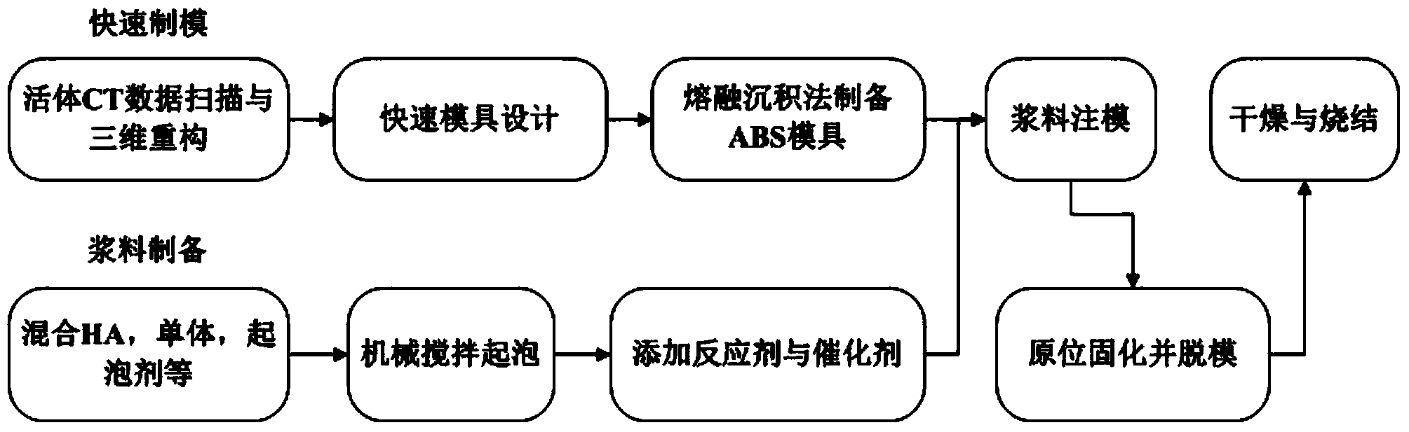
[0046] The preparation method of the personalized porous thyroid cartilage scaffold is as follows:

[0047] Step 1: Use high-precision medical CT to collect data from the living body, and use the segmentation algorithm to segment the thyroid cartilage area, and export the data in text format; the specific method is:

[0048] (I) Use Philips / iCT256 equipment to continuously scan the living body, the scan voltage is 120KV, the scan thickness is 0.625mm, the resolution is 1024*1024, the side length of the field of view window is 140mm, and several two-dimensional tomographic gray images in DICOM format are obtained. These image data are imported into the MIMICS software and stacked in sequence along the helical scanning axis, using the horizontal and vertical axes of the rectangular CT image as the X and Y axes, and the vertical XY plane as the Z axis to establish a spatial rectangular coordinate system;

[0049] (Ii) Using the threshold segmentation algorithm, the threshold is set to 8...

Embodiment 2

[0065] The preparation method of the personalized porous thyroid cartilage scaffold is as follows:

[0066] Step 1: Use high-precision medical CT to collect data from the living body, and use the segmentation algorithm to segment the thyroid cartilage area, and export the data in text format; the specific method is:

[0067] (I) Use Philips / iCT256 equipment to scan the living body continuously, the scan voltage is 125KV, the scan thickness is 0.675mm, the resolution is 1024*1024, the side length of the field of view window is 170mm, and several two-dimensional tomographic gray-scale images in DICOM format are obtained. These image data are imported into the MIMICS software and stacked in sequence along the helical scanning axis, using the horizontal and vertical axes of the rectangular CT image as the X and Y axes, and the vertical XY plane as the Z axis to establish a spatial rectangular coordinate system;

[0068] (Ii) Using the threshold segmentation algorithm, the threshold is se...


PUM


Abstract

The invention discloses a method for preparing an individual porous thyroid cartilage support. The method comprises the following steps: performing data acquisition on a living body by using high-precision medical CT, partitioning a thyroid cartilage area by using a partitioning algorithm, and exporting the data in a text format; performing in-vitro isolated point removal, noise reduction, surface smoothing and reverse three-dimensional modeling on the acquired data in reverse engineering software; performing rapid mold design by using industrial CAD (Computer-Aided Design) software UG-NX, and preparing a rapid mold through rapid molding; preparing foam slurry which takes hydroxylapatite particles as a filling matrix by using a gel foaming method, injecting the slurry into the rapid mold, and initiating an in-situ curing reaction so as to finish preparation of a cartilage support blank; demolding and drying the support blank, and performing high-temperature sintering, thereby obtaining the individual porous thyroid cartilage support. After the support is implanted into the body of a patient, the surrounding cartilage metrocyte and growth factors can be effectively absorbed, a metabolism channel is provided for growth of the cartilage metrocyte, and the cartilage metrocyte grows into individual bones.

Description

Technical field [0001] The invention belongs to the field of individualized and digital design and manufacture of medical implants, in particular to a preparation method of a personalized porous thyroid cartilage stent used for repairing thyroid cartilage defects of the larynx. Background technique [0002] Laryngeal trauma and tumor surgery often bring about large-scale defects of the larynx and trachea, the support of the larynx and trachea cartilage is lost, and the tube cannot be extubated after surgery. Reconstruction and repair of cartilage defects in the larynx, restoration of its supporting function, and maximum retention of breathing, swallowing, and pronunciation functions have important clinical significance. [0003] Bone scaffolds made of bioceramic material hydroxyapatite (HA) have been widely used clinically as bone substitutes because of their inorganic components similar to natural bone components and high biocompatibility. Compared with the dense hydroxyapatite s...

Claims


Application Information

Patent Timeline
Patent Type & Authority Applications(China)
IPC IPC(8): A61L27/56A61L27/42G06F17/50
Inventor 夏薇曹高翔张龙城蒋利浩王灿曾建民
Owner GUANGXI UNIV
Why Eureka
Try Eureka