Human breast cancer cell acetylation p53 protein isotope labeling quantitative method

A technology of human breast cancer cells and isotope labeling, which can be used in measurement devices, material analysis by electromagnetic means, instruments, etc., and can solve problems such as inability to quantify acetylated sites.

Inactive Publication Date: 2014-04-23
CHINA PHARM UNIV
View PDF4 Cites 2 Cited by
  • Summary
  • Abstract
  • Description
  • Claims
  • Application Information

AI Technical Summary

Problems solved by technology

The existing p53 protein quantification method is mainly the Western Blot method, which can only achieve the purpose of relative quantification, and cannot quantify some specific acetylation sites

Method used


Image

  • Human breast cancer cell acetylation p53 protein isotope labeling quantitative method
  • Human breast cancer cell acetylation p53 protein isotope labeling quantitative method
  • Human breast cancer cell acetylation p53 protein isotope labeling quantitative method

Examples


Embodiment 1

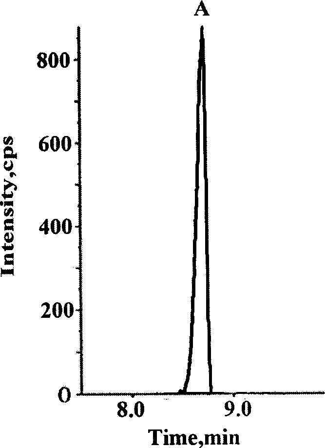
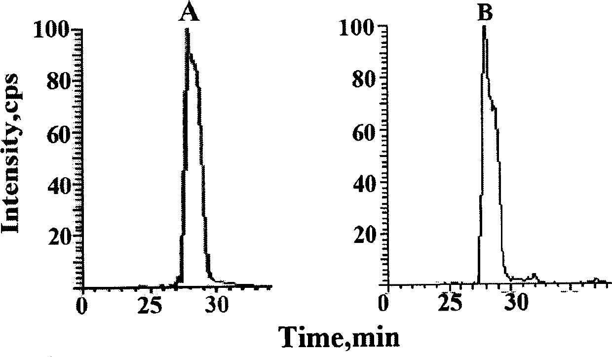
[0034] Example 1: Determination of the amount of acetylated p53 protein in human breast cancer cells at different times after administration of the chemotherapy drug adriamycin

[0035] The main steps are as follows:

[0036] Take the MCF-7 sensitive cells of human breast cancer cells in logarithmic growth phase, digest the cells with 0.1% trypsin, resuspend the cells in complete medium after centrifugation, and count on a blood cell counter to 1×10 4 Each / well was inoculated in a 6-well culture plate. After 24 hours, change the medium after the cells have basically adhered to the wall, and add 200 μl of adriamycin-containing serum-free medium to each well at a concentration of 10 μM. A solvent control administration group (0.1% DMSO) was set for each cell at the same time, and each group had 3 replicate wells. After administration, the cell culture plate is placed in an incubator at 37°C, containing 5% CO 2 , Cultivate in an environment with a relative humidity of 90%. The cells...


PUM


Abstract

The invention belongs to the field of analysis and relates to breast cancer cell endogenous biological macromolecule acetylation p53 protein isotope quantitative analysis. The acetylation p53 protein in a complex cell matrix sample is extracted, separated and purified by utilizing the technical means such as co-immunoprecipitation and gel electrophoresis. The method comprises the following steps: by utilizing a strategy for performing qualitative and quantitative analysis on specific peptide fragments generated by performing trypsin hydrolysis on recombinant human p53 proteins and p53 proteins in breast cancer cells, by taking synthetic peptide fragments generated by performing stable carbon 13 and nitrogen 15 isotope labeling on amino acids in the specific peptide fragment sequence as interior labels, realizing quantitative analysis of acetylation p53 proteins on specific sites in a DNA binding domain in a breast cancer cell sample. The method mainly comprises a sample extraction and gel electrophoresis method, an Obitrap measurement method, preparation of a standard curve and measurement of precision and density. The method is good in linear relationship, high in accuracy and high in reproducibility and can be used for quantitative analysis of acetylation p53 proteins in cells.

Description

Technical field [0001] The invention relates to a quantitative method for acetylating p53 protein at functional sites of DNA binding regions in human breast cancer cells using isotope-labeled binding orbitrap mass spectrometry technology. Background technique [0002] Breast cancer has become the most common cancer among women worldwide. The p53 gene is the tumor suppressor gene with the highest correlation with human tumors. It is closely related to breast cancer tumor growth, apoptosis and drug resistance. After breast cancer cells are stimulated by chemotherapeutic drugs, the expression of p53 protein is up-regulated, and a series of downstream apoptosis programs are initiated, thereby killing tumor cells. The increase in p53 protein expression can significantly inhibit tumor angiogenesis and inhibit tumor metastasis. During DNA damage, p53 undergoes complex post-translational modifications, including phosphorylation, acetylation, methylation, ubiquitination, etc. Among them, ...

Claims


Application Information

Patent Timeline
Patent Type & Authority Applications(China)
IPC IPC(8): G01N27/62
Inventor 王广基汤志远郝海平吴梦秋李盈淳
Owner CHINA PHARM UNIV
Why Eureka
Try Eureka