Non-invasive method for specific 3d detection, visualization and/or quantification of an endogenous fluorophore such as melanin in a biological tissue

A biological tissue and fluorophore technology, applied in fluorescence/phosphorescence, diagnostic recording/measurement, image data processing, etc., can solve problems such as expensive and limited, and achieve rapid implementation, reduce quantity, and improve rapidity

Active Publication Date: 2014-07-16
LOREAL SA
View PDF2 Cites 7 Cited by
  • Summary
  • Abstract
  • Description
  • Claims
  • Application Information

AI Technical Summary

Problems solved by technology

In order to provide a good estimate of the fluorescence lifetime, the FLIM technique requires the integration of the fluorescence signal over time and space, an expensive operation in terms of time, thus limiting the application possibilities of this technique especially in the context of three-dimensional imaging.

Method used


Image

  • Non-invasive method for specific 3d detection, visualization and/or quantification of an endogenous fluorophore such as melanin in a biological tissue
  • Non-invasive method for specific 3d detection, visualization and/or quantification of an endogenous fluorophore such as melanin in a biological tissue
  • Non-invasive method for specific 3d detection, visualization and/or quantification of an endogenous fluorophore such as melanin in a biological tissue

Examples


Embodiment 1

[0093] Example 1: In vivo observation of human skin

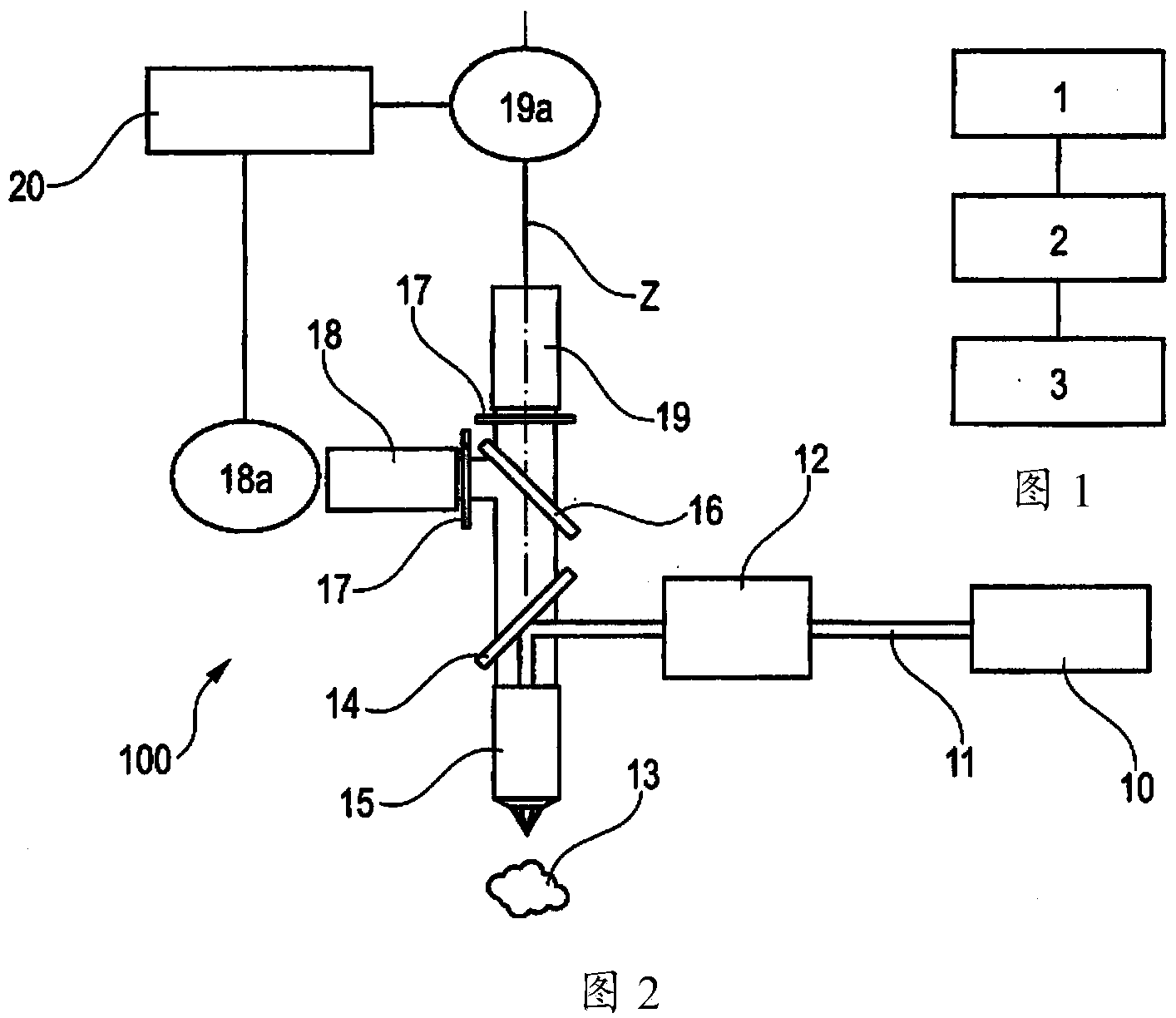
[0094] During the test, three-dimensional images of the human forearm were successively obtained in vivo. One three-dimensional image was composed of a stack of 70 two-dimensional images collected at a depth of 2.346 μm from the surface of the skin, using The device was acquired with an excitation wavelength of 760 nm and an objective lens of 40x / 1.3NA.

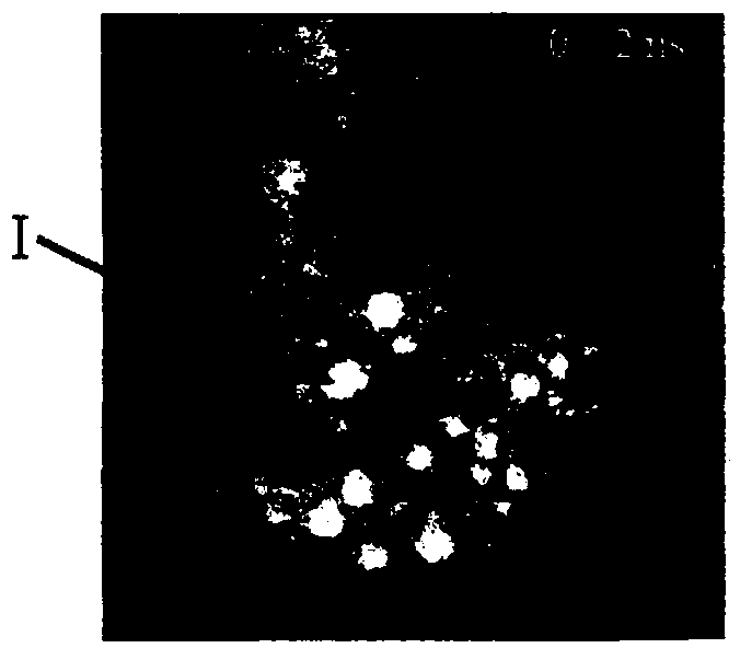
[0095] Figure 3A to Figure 3D A time series of three-dimensional images is shown, in which only four two-dimensional temporal images acquired in vivo on the skin of the forearm of a healthy volunteer, close to the basal layer of the epidermis at a depth of 50 μm relative to the skin surface are shown. For each temporal image, the fluorescence signal was integrated over a period of 2 ns.

[0096] Under experimental conditions, for a spot of a sample containing melanin, most of the emitted fluorescence photons are collected during the first time acquisition. The great...

Embodiment 2

[0103] Example 2: Evaluation of corticosteroid treatment by skin

[0104] On the same area of ​​skin, before and after treatment with corticosteroids by the skin, use as previously described Microscope for observation.

[0105] Figure 9A Raw image showing the fluorescence of a sample of a skin region prior to processing, Figure 9C The corresponding melanin spots obtained by the method according to the invention are shown.

[0106] (In case of occlusion) Treatment of the skin by cutaneous corticosteroids.

[0107] Figure 9B and Figure 9D Raw images of the fluorescence of the samples and the corresponding melanin spots obtained by the method according to the invention after three weeks of treatment are shown.

[0108] The method according to the invention allows the specific detection of melanin; Figure 9C and Figure 9D A comparison of the spots shown in shows a reduction in the amount of melanin, allowing a better assessment of changes caused by cutaneous corti...

Embodiment 3

[0109] Example 3: Characterization of Human Skin Phototype I to Human Skin Phototype IV

[0110] Human skin is classified into six phototypes based on how it reacts to exposure to the sun. Darker skin (phototype V and phototype VI) has a greater amount of melanin, which naturally shields against ultraviolet (UV) rays. The method according to the invention allows a better analysis of pigmentation corresponding to the composition of phototype I to phototype IV of skin ranging from very fair to medium brown.

[0111] Figure 10A and Figure 10B Raw images of fluorescence of skin samples for various phototypes and corresponding melanin spots obtained by the method according to the invention are shown.

[0112] according to Figure 10B spots, calculate the 3D density of melanin in order to obtain Figure 10C , which demonstrates that the amount of melanin increases with light type.

[0113] as passed Figure 10B As shown by the spots corresponding to phototype I, the metho...


PUM


Abstract

Method for detection, quantification and / or visualization of an endogenous fluorophore, in a biological tissue, the method comprising the steps consisting in: a) acquiring, after multiphoton excitation, a set of successive three- dimensional or two-dimensional images providing information on the decrease over time in the fluorescence signals in the sample, b) determining by processing these images, by performing a linear regression of the logarithm of the fluorescence signals, the spatial distribution in the sample of the slopes of said linear regression, and c) generating information on the presence and / or the volume density or surface density of a fluorophore in the sample at least on the basis of this spatial distribution, the fluorophore being melanin.

Description

technical field [0001] The present invention relates to the observation of biological tissues, especially keratinous materials such as skin. [0002] The present invention relates more particularly but not exclusively to the determination of the distribution, especially the quantity and / or the nature, of melanin in biological tissue, especially in order to assess the effect of cosmetic or therapeutic treatments. Background technique [0003] The color of biological tissue is closely related to the quantity and three-dimensional distribution of melanin. Today, the characterization of melanin can be performed in 2D by white-light imaging and quantification of melanin stained by Fontana Masson on histological sections or in 3D by multiphoton imaging and 3D quantification of melanin after image processing. Quantity and distribution. [0004] Multiphoton microscopy facilitates the in-depth observation of biological tissue, especially highly scattering living tissue. This is be...

Claims


Application Information

Patent Timeline
Patent Type & Authority Applications(China)
IPC IPC(8): G01N21/64A61B5/103G06T7/00
Inventor 特蕾泽·巴尔德卫克安娜-玛丽亚·潘纳艾曼纽·坦克雷德-博安艾蒂安·戴西恩赛雷瑟奇·库都罗
Owner LOREAL SA
Why Eureka
Try Eureka