Reagent for mycobacterium tuberculosis infection detection, clinical treatment effect tracking and antituberculous vaccine development and application thereof

A technology for clinical treatment of Mycobacterium tuberculosis, applied in the field of biomedical testing, can solve the problems that specific immunological detection has not been widely used, false positives, unsatisfactory diagnostic value of tuberculosis, etc.

Inactive Publication Date: 2014-08-27
SHANGHAI JIAOTONG UNIV SCHOOL OF MEDICINE
View PDF1 Cites 7 Cited by
  • Summary
  • Abstract
  • Description
  • Claims
  • Application Information

AI Technical Summary

Problems solved by technology

[0004] However, existing studies have shown that ESAT-6, CFP-10 and TB7.7 are mainly expressed in the early infection stage of Mycobacterium tuberculosis, and M.marinum, M.kansasii, M.szulgai or M.gordonae etc. also express ESAT-6, CFP-10 or TB7.7, so false positive results will appear when patients are infected with such...

Method used


Image

  • Reagent for mycobacterium tuberculosis infection detection, clinical treatment effect tracking and antituberculous vaccine development and application thereof
  • Reagent for mycobacterium tuberculosis infection detection, clinical treatment effect tracking and antituberculous vaccine development and application thereof
  • Reagent for mycobacterium tuberculosis infection detection, clinical treatment effect tracking and antituberculous vaccine development and application thereof

Examples


Embodiment 1

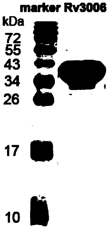
[0077] Example 1: Preparation of Mycobacterium tuberculosis Rv3006 antigen and polypeptide.

[0078] 1. The experimental steps for the preparation of Rv3006 fusion protein (antigen) are as follows:

[0079] 1. Use PCR to amplify the Rv3006 gene from Mycobacterium tuberculosis DNA. After sequencing, insert the gene fragment into the E.coli expression plasmid pET28a to construct the pET28a-Rv3006 prokaryotic expression plasmid.

[0080] 2. The pET28a-Rv3006 prokaryotic expression plasmid was transformed into Escherichia coli E.coli BL21(DE3), and the expression engineered bacteria were constructed.

[0081] 3. Use 0.4mM IPTG at 30℃, 220rpm to induce Rv3006 antigen expression for 3h.

[0082] 4. After ultrasonically lysing the bacteria, centrifuge to collect the supernatant protein solution.

[0083] 5. Purify the Rv3006 fusion protein with Ni-NTA magnetic beads.

[0084] 6. The concentration of the fusion protein was detected by the BCA (Pierce, 23227) method, and stored in a...

Embodiment 2

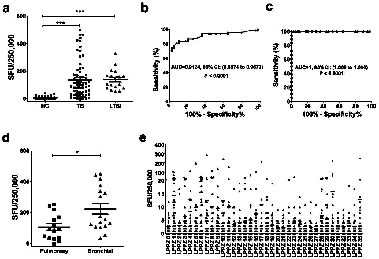
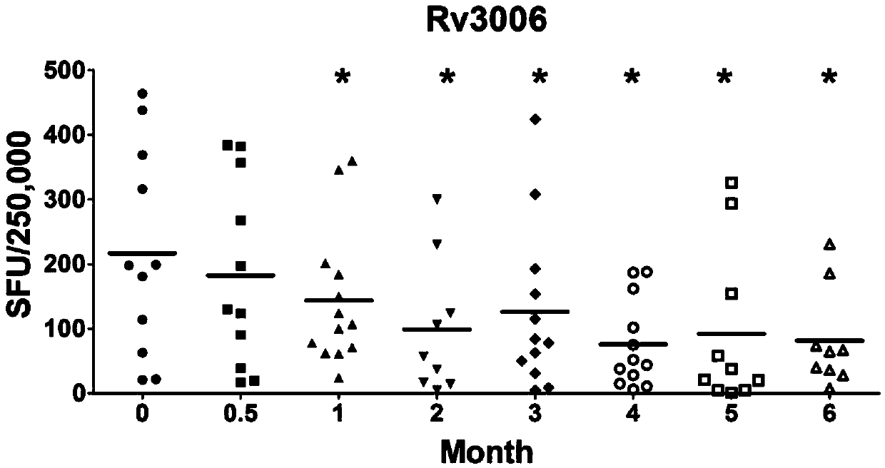
[0124] Example 2: ELISPOT detects the number of specific gamma-interferon-releasing cells stimulated by Rv3006 antigen and its polypeptide

[0125] The experimental steps are as follows:

[0126] 1. Coat ELISPOT 96-well PVDF plate (Millipore MSIPS4510) with IFN-γ antibody, and keep overnight at 4°C.

[0127] 2. Add 200 μl of PBS or RPMI1640 to each well to wash 3 times, and block with RPMI1640 containing 10% FBS at 37° C. for 1 hour.

[0128] 3. Use an anticoagulant tube containing sodium heparin, sodium citrate or dipotassium EDTA to collect about 5 ml from the periphery of tuberculosis patients or normal control populations.

[0129] 4. Using Ficoll density gradient centrifugation method, centrifuge at 1000×g, 18-22°C for 22min. Peripheral blood mononuclear cells (PBMC) were isolated.

[0130] 5. PBMC were washed twice with 5-10ml PBS or RPMI1640 and resuspended in RPMI1640 containing 10% FBS. Calculate the number of cells and adjust to a final concentration of 2.5×10 6 ...

Embodiment 3

[0139] Example 3: ELISA detection of Rv3006-specific antibody levels in the peripheral blood of active tuberculosis patients and normal control populations.

[0140] The experimental steps are as follows:

[0141] 1. 1 μg / ml of Rv3006 fusion protein was coated with ELISA 96-well plate (Corning9018), and kept overnight at 4°C.

[0142] 2. Discard the supernatant, wash each well with 300 μl of PBS (PBST) containing 0.05% Tween-20 5 times, and pat dry each time on absorbent paper.

[0143] 3. Block with PBS containing 1% gelatin at 37°C for 1 hour.

[0144] 4. Add 300 μl PBST to each well to wash 5 times. Add 100 μl of peripheral blood plasma from tuberculosis patients or normal persons diluted 1:100 to each well, and the dilution solution is PBST containing 1% gelatin. Incubate at 37°C for 1 hour.

[0145] 5. Add 300 μl PBST to each well to wash 6 times. Add 100 μl of HRP-labeled anti-human IgG antibody diluted in PBST containing 1% gelatin to each well, and incubate at 37°...


PUM

PropertyMeasurementUnit
Molecular weightaaaaaaaaaa

Abstract

The invention discloses a reagent for mycobacterium tuberculosis infection detection, clinical treatment effect tracking and antituberculous vaccine development. The novel mycobacterium tuberculosis derived reagent Rv3006 (LPPZ) prepared by a gamma-interferon gamma release assay and enzyme linked immunosorbent assay comprises antigen or polypeptide and analogues thereof, wherein terminal C to terminal N in the amino acid sequence of the Rv3006 antigen is as shown in SEQ ID NO:1. By utilizing the reagent, tuberculous patients in the active phase and tuberculous latent infectors can be well detected; the specific immune response of Rv3006 is reduced along with treatment development and disease condition remission, which indicates that the reagent can be used for well tracking the clinical treatment situation of a tuberculous patient, thus having a good clinical application prospect. Furthermore, the reagent has a protective effect on a mouse infected by a mycobacterium tuberculosis standard toxic strain H37Rv, thus having a good development prospect of antituberculous vaccines.

Description

technical field [0001] The invention belongs to the field of biomedical inspection, and in particular relates to a reagent used for detecting mycobacterium tuberculosis infection, tracking the curative effect of clinical treatment and developing an anti-tuberculosis vaccine and its application. Background technique [0002] Tuberculosis is one of the three major infectious diseases in my country and the world. In 2013, the World Health Organization (WHO) estimated that the total number of tuberculosis cases in China was about 1.4 million, accounting for 12% of the total cases in the world, second only to India. The second in the world, tuberculosis seriously hinders my country's economic and social development. [0003] At this stage, the diagnosis and prognosis of tuberculosis and the development of anti-tuberculosis vaccines still face great challenges. Traditional diagnostic methods such as tuberculin skin test (TST), tuberculosis sputum smear or tuberculosis sputum cultu...

Claims


Application Information

Patent Timeline
IPC IPC(8): C07K14/35G01N33/68G01N1/30A61K39/04A61P31/06
CPCC07K14/35A61K39/00G01N33/5695
Inventor 肖洋炯王颖沈浩王慧宇田兆峰赖允鑫
Owner SHANGHAI JIAOTONG UNIV SCHOOL OF MEDICINE
Why Eureka
Try Eureka