
Intracranial tumor operation planning and simulating method based on 3D print technology

A 3D printing and surgical planning technology, applied in the field of medical image processing and application, can solve the problems of inoperable, difficult to operate tumors, short course of malignant tumors, etc., and achieve the effect of simple implementation process, improved accuracy and success rate

Inactive Publication Date: 2014-10-08
刘宇清 +4
View PDF0 Cites 76 Cited by
  • Summary
  • Abstract
  • Description
  • Claims
  • Application Information

AI Technical Summary

Problems solved by technology

[0003] Recently, surgery, chemotherapy, radiotherapy, X-knife, γ-knife, etc. are mostly used in the treatment of intracranial tumors at home and abroad, but most of them are difficult to cure. Malignant tumors have a short course of disease and rapid development. Sensitivity, recurrence may be early or late, tumors growing in important parts such as brainstem and thalamus are difficult to operate or inoperable, tumors after X-knife, radiation and other treatments may shrink or control growth in a short period of time, and then the tumor will no longer grow , Recurrence, benign tumors have a long course of disease and slow growth. Complete surgical resection is not easy to relapse, but brain tumors that grow in important parts such as the brainstem can only be partially or mostly resected, and the tumor will recur after surgery. Growth and intracranial malignant tumors have an average survival rate of less than one year after surgery with radiotherapy and chemotherapy. Benign intracranial tumors such as benign gliomas, pituitary tumors, craniopharyngiomas, and cholesteatomas are not easy to remove completely. Gamma knife, X-ray Knife and radiation therapy cannot completely kill the tumor, so most patients will still relapse after surgery
However, this virtual model lacks an effective physical look and feel, and it is difficult to provide an effective method and platform for comprehensive analysis of comprehensive simulated surgical plans, risks, and prognosis

Method used


Image

  • Intracranial tumor operation planning and simulating method based on 3D print technology
  • Intracranial tumor operation planning and simulating method based on 3D print technology

Examples


Embodiment Construction

[0031] In order to make the above-mentioned features and advantages of the present invention more comprehensible, the following specific embodiments are described in detail with reference to the accompanying drawings.

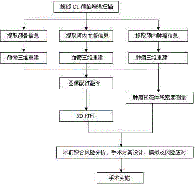
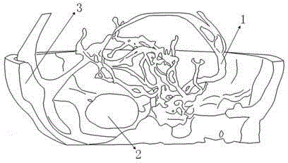
[0032] refer to figure 1 and figure 2 As shown, the 3D printing technology-based simulation method for intracranial tumor surgery planning in the present invention includes the following steps:

[0033] (1) Based on the two-dimensional original image data of the patient's preoperative spiral CT enhanced brain scan, the three-dimensional models of the skull, blood vessels and tumors were reconstructed.

[0034] (2) Measurement of tumor shape, size, density changes and differences. The three-dimensional measurement of tumor shape and size can preliminarily grasp the basic information and characteristics of tumors, and the measurement of tumor density changes and differences can be used as the basis for early diagnosis.

[0035] (3) Registration and fusion of ...


PUM

PropertyMeasurementUnit
Grayscaleaaaaaaaaaa

Abstract

The invention relates to an intracranial tumor comprehensive operation planning and simulating method based on the 3D print technology. DICOM data obtained through patient spiral CT craniocerebral enhancement scanning are utilized first, different partition methods are adopted for data information extraction and reconstruction according to characteristics of skulls, blood vessels and tumors, and registration and fusion of the skulls, the blood vessels and the tumors are achieved in the same coordinate system; craniocerebral entity models of patients are obtained through a rapid prototyping technology and a virtual three-dimensional geometrical model after optimizing processing is conducted; information of diameters, sizes and the number of tumors and adjacent relations between the diameters, the sizes and the number of tumors and important blood vessels and tissue around is extracted, and preoperative comprehensive risk analysis, operation plan design, operation plan simulation and operation risk response analysis are carried out on the basis of the craniocerebral entity models of the patients, personalized operation plans are provided for the patients in order to improve the accuracy and success rate of intracranial tumor excision operations, and a feasible intracranial tumor operation planning and simulating method based on the 3D print technology is explored.

Description

technical field [0001] The invention relates to a method for planning and simulating intracranial tumor surgery based on 3D printing technology, and belongs to the field of medical image processing and application. Background technique [0002] Intracranial tumors, namely various brain tumors, are one of the common diseases in the nervous system, which have great harm to the function of the human nervous system. In recent years, the incidence of intracranial tumors has been on the rise. According to statistics, intracranial tumors account for about 5% of systemic tumors and 70% of childhood tumors, while 20-30% of other malignant tumors will eventually transfer to the brain. Once its expanding and infiltrating growth occupies a certain space in the brain, no matter whether its nature is benign or malignant, it will inevitably increase intracranial pressure, compress brain tissue, cause central nervous system damage, and endanger the patient's life. [0003] Recently, surger...

Claims


Application Information

Patent Timeline
IPC IPC(8): G06T7/00G06T17/00A61B6/03
Inventor 刘宇清姚立纲黄绳跃何炳蔚洪文瑶
Owner 刘宇清
Learn More