Method for detecting degradation of protein in cells

A protein and cell technology, applied in the field of biochemistry, can solve the problems of high laboratory requirements, blocking, high cytotoxicity of cycloheximide, etc.

Inactive Publication Date: 2014-11-19
FUDAN UNIV
View PDF4 Cites 4 Cited by
  • Summary
  • Abstract
  • Description
  • Claims
  • Application Information

AI Technical Summary

Problems solved by technology

Practice has shown that the main defects of this classic protein degradation detection method are as follows: 1. It needs to use isotopes, so it has a certain impact on the health and safety of experimenters and has high requirements for the laboratory; 2. The operation process is complicated and requires High-throughput incompatible experimental steps such as immunoprecipitation and electrophoretic separation make it impossible to use this method for high-throughput screening for protein degradation; Quantification of rates is noisy and imprecise in many cases
[0004] In recent years, some studies have used the protein translation blocker cycloheximide to study protein degradation, which has the advantages of convenient operation and the possibility of high-throughput screening; however, there are still the following defects: 1. Cycloheximide blocks all proteins Translation, strong non-specificity; 2. Cycloheximide has high cytotoxicity, and it will cause large-scale cell death after 9 hours of treatment. Therefore, it is not suitable for proteins with slower degradation rates; on the other hand, in fact many Slow protein degradation in neurodegenerative diseases
However, the operating procedure used is similar to the traditional pulse-trace experiment, except that the azide small molecule group is used instead of the isotope 35 S markers, therefore, still cannot achieve high-throughput and accurate quantification

Method used


Image

  • Method for detecting degradation of protein in cells
  • Method for detecting degradation of protein in cells

Examples


Embodiment 1

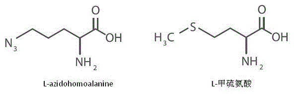
[0031] Example 1 Methionine Labeling of Newly Synthesized Proteins in Cells by Azide Small Molecule Groups

[0032] In various cells such as STHdh of experimental mice, methionine is an essential amino acid for protein synthesis. L-azidohomoalanines are methionine analogues with an azido group. Spread the same number of cells in several culture dishes, culture with complete culture medium for 24 hours, remove the complete culture medium, and wash once with PBS. The cells were starved for one hour using the methionine-free medium purchased from Life Technologies to completely consume the original methionine in the cells, and then the azido-containing methionine analog L -azidohomoalanine, so that the final concentration in the culture medium is 1‰, culture for 12 hours, during the 12 hours, the newly synthesized protein in the cells is labeled with L-azidohomoalanine, so as to have an azide group; collect the first time point after 12 hours Wash the cells in the remaining cul...

Embodiment 2

[0034] The biotin molecule is coupled to the azide small molecule group of the protein by CuAAC reaction:

[0035] The CuAAC reaction refers to the cycloaddition reaction of copper catalyzed azide and terminal alkyne, using copper ions to catalyze the reaction of the azide group of the protein that has been labeled with the azide group but not degraded with the terminal alkyne with biotin , to achieve protein labeling with biotin.

[0036] The reaction was carried out according to the specific operation steps of the Click-iT Reaction Kit purchased from Life Technologies. Add up to 50uL of protein lysate to the EP tube (up to 200ug of protein), add 100uL of Click-iT reaction buffer, add water to make up to a total volume of 160uL, Vortex for 5s, add 10uL of Click-iT Component B (CuSO 4 ), Vortex for 5s, add 10uL click it reaction buffer additive 1, Vortex for 5s, place for 2-3 min, but no more than 5 min, add 20uL Click-iT reaction buffer additive 2, Vortex for 5s, place t...

Embodiment 3

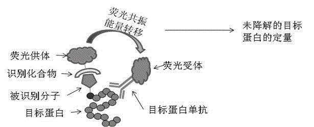
[0038] Quantification of target proteins coupled with biotin molecules using the principle of homogeneous time-resolved fluorescence resonance:

[0039] Streptavidin (Streptavidin) and biotin (Biotin) have specific binding properties, adding fluorescent group-labeled streptavidin and target protein antibody in the reaction, using the principle of homogeneous time-resolved fluorescence resonance ( HTRF), to perform high-throughput, high-precision, and specific quantification of undegraded but labeled target proteins, thereby accurately detecting protein degradation.

[0040] Add a certain amount of biotin-labeled protein samples into a 384-well plate, add Streptavidin-D2 labeled with fluorescent receptor D2 (1.4 ng / μl ) and Terbium Cryptate-labeled target protein-specific antibody (2B7-Tb, 0.023 ng / μl), after incubation for 1 to 2 hours, detect fluorescence resonance signals on a PerkinElmer Envision instrument, only those labeled with biotin , and the protein that can be spec...


PUM


Abstract

The invention belongs to the field of biochemistry, and relates to a method for detecting the degradation of protein in cells. The method comprises the following steps: (1) marking methionine of newly synthesized protein in the cells with azide small molecule groups by utilizing an methionine analogue; (2) lysing the cells at different time points, and coupling specific molecules which can be identified to the azide small molecule groups through CuAAC reaction; and (3) quantifying target protein which is coupled with the molecules which can be identified in the step (2) by directly utilizing an HTRF (Homogeneous Time-Resolved Fluorescence) resonance principle in a reaction system so as to determine the degradation rate of the target protein. The method is a new protein degradation measurement method which is free from the need of an isotope or cytotoxin, is compatible with high-throughput screening, and can be used for screening related disease protein degradation medicines in pharmaceutical industry, screening specific related protein degradation signals in academia, and the like.

Description

technical field [0001] The invention belongs to the field of biochemistry and relates to a method for detecting intracellular protein degradation, in particular to a detection method for intracellular protein degradation based on CuAAC reaction and energy transfer homogeneous time-resolved fluorescence resonance. Background technique [0002] It is well known in the art that protein degradation and its related parameters (degradation rate and half-life, etc.) are of great significance for understanding protein function, the expected effect of small molecule regulators on protein, and developing drugs for the degradation of specific disease proteins. At present, the protein degradation pathways reported mainly include proteasomal degradation and lysosomal degradation. Studies have shown that when proteins are degraded abnormally through the above two pathways, diseases may occur. For example, a variety of neurodegenerative diseases (such as Huntington's disease, etc.) are ca...

Claims


Application Information

Patent Timeline
Patent Type & Authority Applications(China)
IPC IPC(8): G01N33/68G01N21/64
CPCG01N33/54373G01N33/582G01N33/6872
Inventor 鲁伯埙崔笑添
Owner FUDAN UNIV
Why Eureka
Try Eureka