Improved stem/progenitor cell and regenerative porcine islet cell co-culturing method
A technology of co-cultivation of islet cells, applied in blood/immune system cells, bone/connective tissue cells, animal cells, etc., can solve the problem of not involving newborn pig islet cells, low efficiency, complicated procedure of stem cell encapsulation of islet cells, etc. problems, to achieve the effect of being suitable for transplantation, increasing efficiency, and high coating efficiency
- Summary
- Abstract
- Description
- Claims
- Application Information
AI Technical Summary
Problems solved by technology
Method used
Image
Examples
Embodiment 1
[0029] Example 1. Separation and purification of neonatal porcine islet cells
[0030]1. The experiment was carried out in accordance with the requirements of the 2006 "Guiding Opinions on Treating Experimental Animals" issued by the Ministry of Science and Technology. Newborn pigs born 3-5 days old were adequately anesthetized with 3% pentobarbital, and the pancreas was taken by sterilized laparotomy. Place the removed pancreatic tissue in a pre-cooled D-Hanks liquid glass dish, remove the capsule, fat and blood vessels around the pancreatic tissue block, weigh them, and then cut the pancreatic tissue block into a tissue size of about 0.5-1mm3 piece.
[0031] 2. Add an appropriate amount of type V collagenase, place in a 37°C water bath for shaking digestion, and obtain islet cell suspension.
[0032] 3. Add the islet cell suspension to the culture dish with complete medium (FAMS / F-10, containing 10% porcine serum, streptomycin 100U / mL, penicillin 100U / mL, 0.1% glutamine) ...
Embodiment 2
[0033] The cultivation and identification of embodiment 2.MSCs
[0034] 1. Subculture of MSCs
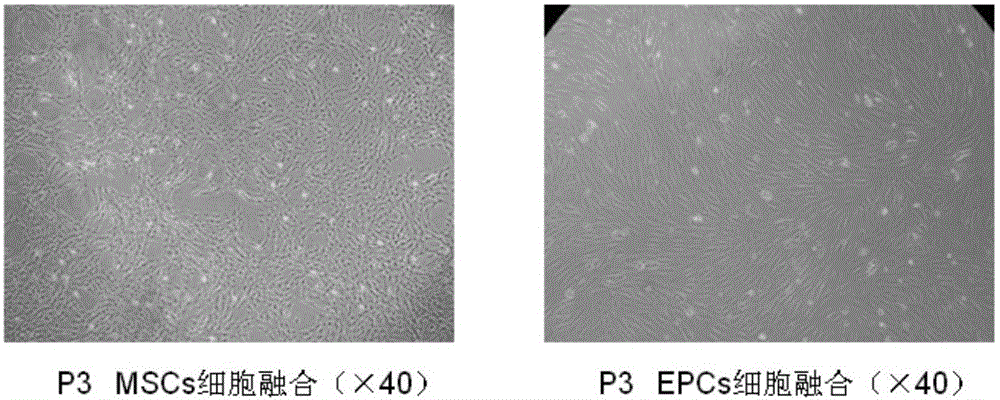
[0035] The isolated and purified umbilical cord-derived mesenchymal stem cells were resuscitated from the liquid nitrogen tank, cultured with MSC medium, and the medium was changed every three days, and the culture was continued until 4-5 days. It can be seen that the cells are more than 90% confluent. 1x10 4 cells / cm 2 (2.5x10 5 cells / T25 culture flask) for subculture, and the P3-5 passage MSCs were taken for experiments.
[0036] 2. Identification of MSCs
[0037] Take the umbilical cord-derived mesenchymal stem cells of the P3-5 generation, remove the medium, wash twice with PBS, and digest with trypsin, stop the digestion with medium containing serum, wash with PBS, resuspend the cells, and adjust the cell concentration to 1×10 6 / mL single cell suspension, take the flow tube and add 0.1ml cell suspension to each tube, add mouse anti-human monoclonal antibodies FITC-CD34, ...
Embodiment 3
[0039] The cultivation and identification of embodiment 3.EPCs
[0040] 1. Subculture of EPCs
[0041] The isolated and purified umbilical cord blood-derived endothelial progenitor cells were resuscitated from the liquid nitrogen tank, inoculated into a culture dish covered with a special layer (such as fibronectin), and cultured by adding special growth factors (such as vascular endothelial growth factor). factor, epidermal growth factor, basic fibroblast growth factor, hepatocyte growth factor, etc.) in EGM-2 medium, cultured for 4-5 days, cell colonies appeared in the adherent cells. 8-10 days, more than 90% cell confluence can be seen, according to 5x10 4 cells / cm 2 (10 6 cells / T25 culture flask) for subculture, and P3-5 generation EPCs for experiments ( figure 1 ).
[0042] 2. Identification of EPCs
[0043] Take cord blood-derived endothelial progenitor cells of P3-5 generation, remove the medium, wash twice with PBS, digest with trypsin, stop digestion with medium...
PUM
 Abstract
Description
Claims
Application Information
 - R&D
- Intellectual Property
- Life Sciences
- Materials
- Tech Scout
- Unparalleled Data Quality
- Higher Quality Content
- 60% Fewer Hallucinations
Browse by: Latest US Patents, China's latest patents, Technical Efficacy Thesaurus, Application Domain, Technology Topic, Popular Technical Reports.
© 2025 PatSnap. All rights reserved.Legal|Privacy policy|Modern Slavery Act Transparency Statement|Sitemap|About US| Contact US: help@patsnap.com


