Method for culturing autologous peripheral blood lymphocyte

A technology of lymphocytes and culture methods, applied in the field of CIK culture of autologous peripheral blood lymphocytes, which can solve the problems of increased expression and does not take into account the effect of T regulatory cell content on tumor immune escape, so as to achieve the effect of improving killing activity

Inactive Publication Date: 2015-02-25
ADLAI NORTYE BIOPHARMA CO LTD
View PDF2 Cites 19 Cited by
  • Summary
  • Abstract
  • Description
  • Claims
  • Application Information

AI Technical Summary

Problems solved by technology

[0005] Insufficiency of the current CIK technology: In the tumor microenvironment, the expression of CTLA4 and PD-1 on the surface of T cells increases, and they bind to the corresponding ligands on APC, th

Method used


Image

  • Method for culturing autologous peripheral blood lymphocyte
  • Method for culturing autologous peripheral blood lymphocyte
  • Method for culturing autologous peripheral blood lymphocyte

Examples


Embodiment 1

[0074] Example 1 A method for extracting peripheral blood mononuclear cells

[0075] A method for extracting peripheral blood mononuclear cells, comprising the steps of:

[0076] (1) Collect 30-100mL of peripheral blood with a sodium citrate anticoagulant tube, transfer the peripheral blood to a 50mL centrifuge tube, centrifuge at 2000rmp for 15min;

[0077] (2) Discard the upper layer of plasma, dilute the blood cells with normal saline so that the ratio of blood cells: normal saline is 1:1, and mix well;

[0078] (3) Pipette 15mL of Ficoll lymphocyte separation solution into a 50mL centrifuge tube, and add the mixed cell suspension evenly and slowly to the upper layer to form a complete interface;

[0079] (4) 1600rpm, centrifuge for 20min, obvious stratification can be seen;

[0080] (5) Collect interface mononuclear cells and wash them twice with PBS;

[0081] (6) Resuspend the cells in PBS and count them for later use.

Embodiment 2

[0082] Example 2 A method for cultivating CIK cells

[0083] A method for cultivating CIK cells, comprising the steps of:

[0084] (1) Use X-VIVO15 serum-free medium to adjust the cell concentration of the mononuclear cells isolated in Example 1 to 1*10 6 pcs / mL, placed at 37°C, 7.5%CO 2 incubator cultivation;

[0085] (2) On the third day, add X-VIVO15 serum-free medium to 100mL, and add 1000U / mL IL-2;

[0086] (3) On the fourth day, add X-VIVO15 serum-free medium to 240mL, and add IL-2 to keep the concentration unchanged;

[0087] (4) On the seventh day, put the cell suspension into a 1.8L culture bag, add X-VIVO serum-free medium to 1000mL, add IL-2 to keep its concentration at 1000U / mL, and add CTLA-4mAb 100ng / mL at the same time mL, PD-1mAb 100ng / mL;

[0088] (5) On the 14th day, the cultured CIK cells were collected by centrifugation and washed twice with 0.9wt% sodium chloride solution. Dilute sodium chloride solution to 100mL.

Embodiment 3

[0089] Example 3 Detection of immunophenotype of CIK cells

[0090] The immunophenotype detection of CIK cells includes the following steps:

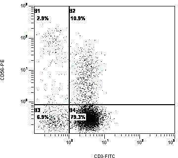
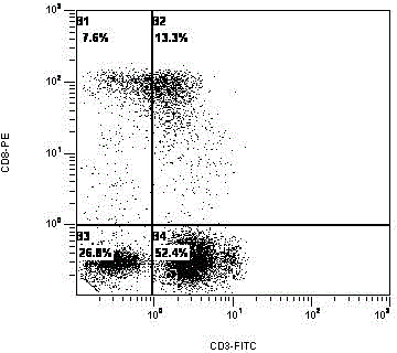
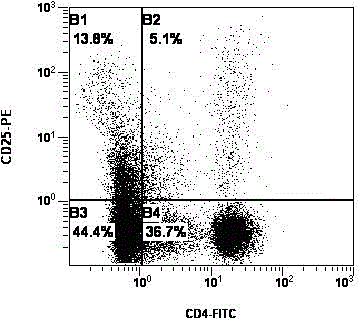
[0091] (1) Take the cells on the 1st, 7th, and 14th day of culture respectively, transfer them into the flow detection tube, add 3mL PBS to mix well, and centrifuge to wash the cells (centrifugation condition: 1600rpm, 5min). Pour off the centrifuged supernatant, according to the concentration and volume of the cell sample, suspend the cells with an appropriate amount of PBS, and adjust the cell concentration to 1x10 5 a / ml;

[0092] (2) Fluorescence-labeled cells: label flow detection tubes, label 4 tubes for each sample, tube 1: IgG-FITC / IgG-PE, tube 2: CD3-FITC / CD56-PE, tube 3: CD3-FITC / CD8-PE, tube 4: CD4-FITC / CD25-PE, add the corresponding fluorescent antibody to each tube, the volume of each antibody is 8 μL, add 100ul of the cell sample suspension to be tested in each tube;

[0093] (3) Gently rotate and mix on a vortex shake...


PUM


Abstract

The invention relates to a method for culturing an autologous peripheral blood lymphocyte. The method comprises the following steps: (1) separating a mononuclear cell from peripheral blood, resuspending in an X-VIVO15 serum-free culture medium to obtain cell concentration of 1*10<6>/mL, and culturing for 3 days; (2) supplementing the X-VIVO15 serum-free culture medium to 100 mL, adding IL-21*10<3> U/mL, and culturing for 1 day; (3) supplementing the X-VIVO15 serum-free culture medium to 200-240 mL, adding IL-21*10<3> U/mL, and culturing for 3 days; (4) supplementing the X-VIVO15 serum-free culture medium to 1000 mL, adding IL-21*10<3> U/mL, CTLA-4mAb100n g/mL and PD-1mAb100n g/mL; and (5) culturing for 5-7 days to prepare the autologous peripheral blood lymphocyte. The method disclosed by the invention can be used for improving the activation efficiency and amplification efficiency of an effector cell group by adding multiple monoclonal antibodies and cell factors to the X-VIVO15 serum-free culture medium, and can be used for effectively reducing the content of T regulatory cells by covering CTLA-4 and PD-1 molecules of the surfaces of all CIK cells by loading CTLA-4 and PD-1 antibodies in vitro especially, thus further enhancing the killing effect of the CIK cells on tumors.

Description

technical field [0001] The invention belongs to the technical field of in vitro culture of immune cells, in particular to a method for culturing autologous peripheral blood lymphocyte CIK. Background technique [0002] Adoptive immunotherapy is to infuse sensitized lymphocytes (with specific immunity) or their products to people with low cellular immunity (such as tumor patients) to obtain anti-tumor immunity. It is an immune system used to treat tumors. therapy. [0003] Cytokine induced killer cells (CIK) are human peripheral blood mononuclear cells co-cultured with various cytokines (such as anti-CD3 monoclonal antibody, IL-2 and IFN-γ, etc.) for a period of time in vitro A population of heterogeneous cells obtained later. Because this kind of cell expresses both CD3+ and CD56+ membrane protein molecules, it is also called NK cell-like T lymphocyte, which has both the strong anti-tumor activity of T lymphocytes and the non-MHC-restricted tumor killing advantages of NK c...

Claims


Application Information

Patent Timeline
IPC IPC(8): C12N5/0783
Inventor 何学松何南海叶晓峰林晓峰杨东晖路杨
Owner ADLAI NORTYE BIOPHARMA CO LTD
Why Eureka
Try Eureka