
Aptamer and manganese porphyrin catalysis-based chemiluminescence protein detection method

A chemiluminescence detection and nucleic acid aptamer technology, applied in the detection field, can solve the problems of poor stability, high cost, cumbersome steps, etc., and achieve the effects of easy synthesis, low test cost and simple operation.

Active Publication Date: 2015-05-20
CHANGCHUN INST OF APPLIED CHEMISTRY - CHINESE ACAD OF SCI
View PDF4 Cites 2 Cited by
  • Summary
  • Abstract
  • Description
  • Claims
  • Application Information

AI Technical Summary

Problems solved by technology

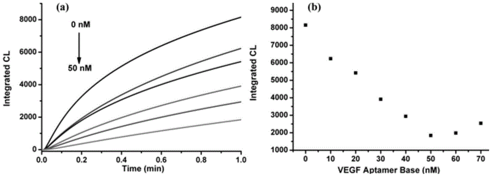
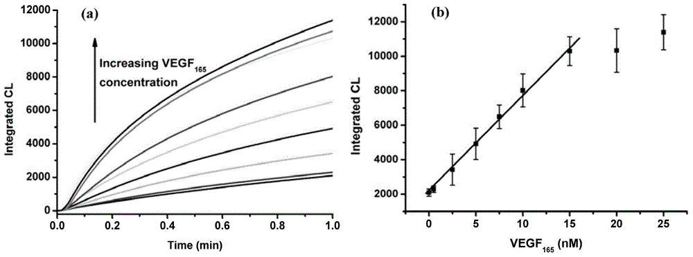
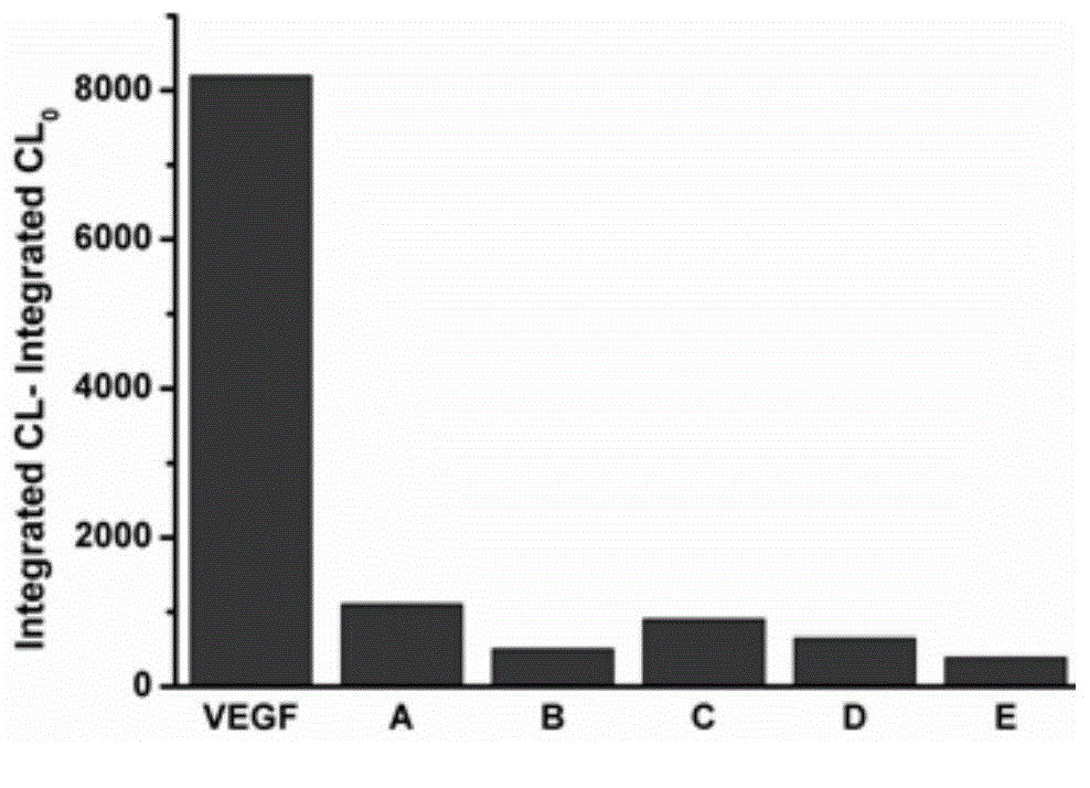
[0004] The present invention aims to solve the technical problems of low sensitivity, poor stability, cumbersome steps, and high cost in the prior art protein detection method based on nucleic acid aptamers, and provides a method based on nucleic acid aptamers and manganese porphyrin catalysis A method for detecting proteins by chemiluminescence, which is a rapid, sensitive, specific and simple protein detection method based on the aggregation of manganese porphyrins induced by nucleic acid aptamers

Method used


Image

  • Aptamer and manganese porphyrin catalysis-based chemiluminescence protein detection method
  • Aptamer and manganese porphyrin catalysis-based chemiluminescence protein detection method
  • Aptamer and manganese porphyrin catalysis-based chemiluminescence protein detection method

Examples


Embodiment 1

[0035] (1) Preparation of manganese porphyrin probe

[0036] Add 5,10,15,20-tetrakis(1-methyl-4-pyridyl)porphyrin (TMPyP) 101mg, MnCl 2 4H 2 O 16mg was dissolved in 10mL of methanol, heated to reflux under nitrogen protection, and the UV-visible spectrum of the reaction solution was monitored. The maximum absorption peak of the Soret absorption band of porphyrin moved from 422nm to 463nm, indicating that porphyrin and manganese have been coordinated. After the completion of the coordination reaction, it was exchanged with a chloride ion exchange resin for 24 hours, then precipitated with ethyl acetate, filtered, and the obtained purple-black solid was dried in a vacuum oven to prepare a manganese porphyrin probe (MnTMPyP). The synthetic route is as follows:

[0037]

[0038] (2) VEGF 165 detection

[0039]Add 970 μL of buffer solution (the buffer solution contains 20 mM Tris-HCl, 25 μM luminol, 0.05% Triton X-100) to a small glass beaker, then add 2 μL of 5 μM MnTMPyP,...

Embodiment 2

[0041] (1) Preparation of manganese porphyrin probe

[0042] Add 5,10,15,20-tetrakis(1-methyl-4-pyridyl)porphyrin (TMPyP) 101mg, MnCl 2 4H 2 O 16mg was dissolved in 10mL of methanol, heated to reflux under nitrogen protection, and the UV-visible spectrum of the reaction solution was monitored. The maximum absorption peak of the Soret absorption band of porphyrin moved from 422nm to 463nm, indicating that porphyrin and manganese have been coordinated. After the completion of the coordination reaction, it was exchanged with a chloride ion exchange resin for 24 hours, then precipitated with ethyl acetate, filtered, and the obtained purple-black solid was dried in a vacuum oven to prepare a manganese porphyrin probe (MnTMPyP). The synthetic route is as follows:

[0043]

[0044] (2) Detection of PDGF

[0045] Add 970 μL of buffer solution (the buffer solution contains 20 mM Tris-HCl, 25 μM luminol, 0.05% Triton X-100) to a small glass beaker, then add 2 μL of 5 μM MnTMPyP, ...


PUM


Abstract

The invention relates to an aptamer and manganese porphyrin catalysis-based chemiluminescence protein detection method, which aims at solving the technical problems that an aptamer-based protein detection method is low in sensitivity, poor in stability, complicated in steps, high in cost and the like. The method comprises the following steps: (1) preparing a manganese porphyrin probe; and (2) detecting protein, namely adding 20mM Tris-HCl, 25microM luminol and 0.05% Triton X-100 into a chemical reactor, then adding the manganese porphyrin probe, finally, adding a solution containing an aptamer and the to-be-detected protein, placing the chemical reactor into a chemiluminescence detector, finally, adding 1000mM H2O2 with a trace injector, and recording the change of chemiluminescence along time. The method is an aptamer induction-based fast, sensitive, specific and simple protein detection method which is established by gathering manganese porphyrin.

Description

technical field [0001] The invention relates to a detection method, in particular to a method for detecting protein based on nucleic acid aptamer and chemiluminescence catalyzed by manganese porphyrin. Background technique [0002] Protein is the material basis of life. Without protein, there would be no life. It is closely related to various forms of life activities. The quantitative and analytical detection of protein plays a very important role in basic research and clinical application. At this stage, in the fields of medical diagnosis and analytical chemistry, the most common method for detecting proteins is still based on the specific interaction of antigen-antibody. Due to the strong affinity between antigen and antibody, the detection method has high sensitivity and good specificity. However, the detection method based on immunity also has its disadvantages: the screening, identification, and isolation process of antibodies depend on animal and cell culture, and th...

Claims


Application Information

Patent Timeline
IPC IPC(8): G01N21/76C07D487/22
Inventor 于聪李文英张青峰陈健周会鹏李永新
Owner CHANGCHUN INST OF APPLIED CHEMISTRY - CHINESE ACAD OF SCI
Learn More