Method for changing ultrastructure of cell by using focused ultrasound

A focused ultrasound and ultrastructural technology, applied in the fields of biology and ultrasound, can solve the problems of cell lysis, inability to accurately locate, affect cell channel conduction, etc., and achieve the effect of reducing damage.

Inactive Publication Date: 2015-07-15
JIANGSU PROVINCIAL HOSPITAL OF TCM
View PDF0 Cites 4 Cited by
  • Summary
  • Abstract
  • Description
  • Claims
  • Application Information

AI Technical Summary

Problems solved by technology

[0005] Aiming at the problems existing in changing the ultrastructure of cells in the prior art: (1) incapable of precise positioning; (2) easy to cause cell lysis; (3) electrical stimulation may affect the conduction of cell channels, etc., the present invention provides a method using Method of Focused Ultrasound to Change Cell Ultrastructure

Method used


Image

  • Method for changing ultrastructure of cell by using focused ultrasound
  • Method for changing ultrastructure of cell by using focused ultrasound
  • Method for changing ultrastructure of cell by using focused ultrasound

Examples


Embodiment 1

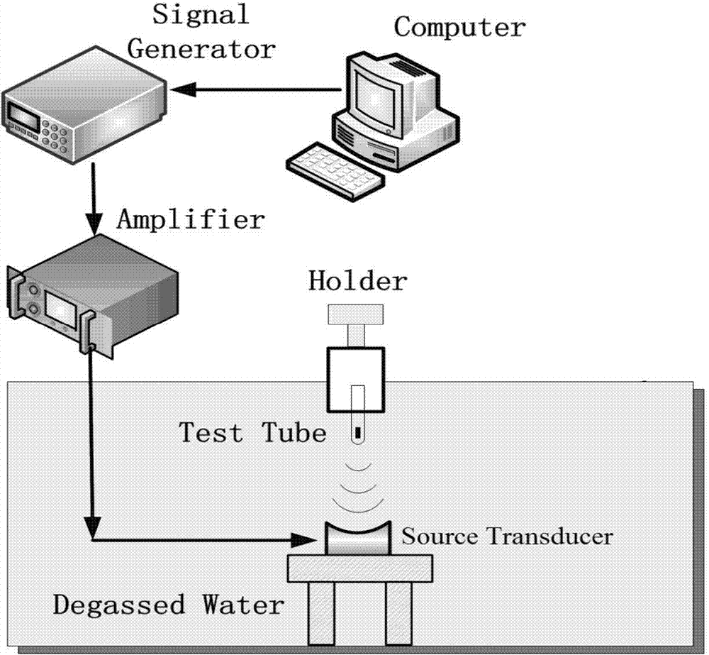
[0033] to combine figure 1 , a method of using focused ultrasound to change the ultrastructure of cells in this embodiment, the specific steps are as follows:

[0034] Step 1. Establishment of cerebral ischemia-reperfusion cell injury model:

[0035] a. Determine the glutamate concentration and action time for establishing the cell injury model. The specific determination process is as follows: take PC12 cell lines, treat them with nerve growth factor, grow and differentiate synapses; then treat the cells with DMEM medium containing different concentrations of glutamic acid, and the selected glutamic acid concentrations are 50, 25, 12.5 , 6.25umol / L, the selected time points were 12, 24, and 48 hours, and then the apoptosis rate of PC12 cells was detected by flow cytometry, and the optimal concentration and treatment for establishing a glutamate model were selected according to the cell apoptosis time. In the course of the experiment, we found that when the concentration o...


PUM


Abstract

The invention discloses a method for changing the ultrastructure of a cell by using focused ultrasound, which belongs to the fields of biology and ultrasonics. The method comprises the following steps: (1) carrying out ultrasonic cavitation on the cell: a, digesting and treating the cell with pancreatin and discarding supernatant; b, adding a DMEM medium and micro bubbles of an ultrasonic contrast medium and putting an obtained cell mixed liquid in a sealed tube; and c, employing a coaxial focused ultrasound transmitting apparatus, placing a focused ultrasound energy transducer at the bottom of the apparatus and installing the sealed tube filled with the cell mixed liquid on a fixator located at the top of the apparatus, wherein the focused ultrasound energy transducer and the sealed tube are guaranteed to be coaxial and the distance between the focused ultrasound energy transducer and the sealed tube can be continuously adjusted; and (2) observing changing of the surface microstructure of the cell strain under the action of ultrasound by using an electron microscope. The method can increase gaps in the surface of the cell through the ultrasonic cavitation effect.

Description

technical field [0001] The invention relates to the fields of biology and ultrasound, and relates to a method for changing cell ultrastructure by using ultrasound technology. Background technique [0002] Ultrasonic transfection technology, which uses ultrasonic cavitation effect to cause cell membrane sonication to promote drug delivery and absorption in the lesion area, is also a key topic in the field of ultrasonic therapy at home and abroad. Since the liquid medium or muscle tissue usually contains many tiny gas nuclei or cavities that are invisible to the naked eye, when the ultrasonic wave propagates in it and the sound pressure exceeds a certain threshold, these gas nuclei will expand in the negative sound pressure phase of the sound wave Cavitation bubbles are formed. Under the action of ultrasound, these cavitation bubbles will produce a series of dynamic processes such as oscillation, expansion, contraction and implosion. The currently clinically used ultrasound ...

Claims


Application Information

Patent Timeline
Patent Type & Authority Applications(China)
IPC IPC(8): C12N13/00
Inventor 张春兵滕凤猛
Owner JIANGSU PROVINCIAL HOSPITAL OF TCM
Why Eureka
Try Eureka