Iba1 and LAG-3 dual-gene co-expression recombinant adenovirus vector and preparation method and application thereof
A technology of LAG-3 and recombinant adenovirus, which is applied in the field of preparation of the carrier, can solve the problems of hindering T cell delayed hypersensitivity, many influencing factors, and uneven gene copy of cells, so as to achieve good development and application prospects, inhibit Inflammation response, effect of improving nerve function
- Summary
- Abstract
- Description
- Claims
- Application Information
AI Technical Summary
Problems solved by technology
Method used
Image
Examples
Embodiment 1
[0030] Embodiment 1, the preparation of recombinant adenovirus Ad-LAG-3 / Iba1
[0031] 1. Cloning the full-length cDNA of LAG-3
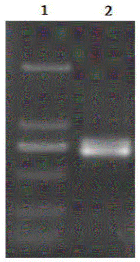
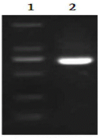
[0032] According to the instructions of the Trizol kit, the total RNA of human liver tissue was extracted, and the total cDNA was obtained by reverse transcription, and then the total cDNA was used as a template, and F1:5'-gg ctcgagg atgtgggaggctcagttcctg-3' (SEQ ID No.1, the underlined part is the Xho Ⅰ restriction site) and R1: 5'-gg gaattctcagagctgctccggctc-3' (SEQ ID No.2, the underlined part is the EcoR Ⅰ restriction site) was used as the upstream and downstream primers to amplify the full-length cDNA of LAG-3 by PCR. The PCR reaction system was 50 μl, and the cycle condition parameters were: Denaturation for 5 minutes; then denaturation at 94°C for 50 seconds, annealing at 58°C for 50 seconds, and extension at 72°C for 2 minutes, a total of 30 cycles; finally, extension at 72°C for 10 minutes. The PCR products were identified by agarose gel...
Embodiment 2
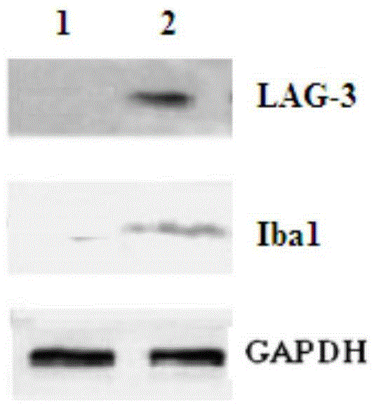
[0044] Example 2, Preparation of recombinant adenovirus Ad-LAG-3 / Iba1 transfected microglial cells
[0045] 1. Sorting of microglia
[0046] Microglial cells were cultured according to the literature method (Lehnart et al, J Neurosci, 2002; 22(7): 2478-2486, this method is a classic microglial cell culture method, widely used in research). The details are as follows: Take the forebrain tissue of C57BL / 6 mice with a reproductive age of 1-2 days, digest with insulin, grind at 37°C for 20 minutes, transfer the cells to DMEM medium containing calf serum for culture, and shake at 180 rpm for 30 minutes after 1 week. The cell suspension (containing >90% microglial cells) was transferred to an uncoated culture plate, and the non-adherent cell suspension was removed after 15 minutes, washed with PBS for 3 times, and the purity of the cells was identified by immunostaining (90% small glial cells) Glial cells).
[0047] 2. Recombinant adenovirus Ad-LAG-3 / Iba1 transfected microglial ce...
Embodiment 3
[0050] Example 3, Anti-proliferation detection of microglial cells transfected with recombinant adenovirus Ad-LAG-3 / Iba1
[0051] The microglial cells were randomly divided into two groups: the experimental group and the control group. The microglial cells in the experimental group were transfected with Ad-LAG-3 / Iba1 virus, and the control group were cultured normally.
[0052] 1. Detection of the proliferation ability of microglial cells
[0053] Adjust the cell concentration to 1×10 with RPMI 1640 medium containing 10% fetal bovine serum 6 cells / ml, inoculate into 96-well plate, 100 μl per well, set 3 duplicate wells for each group, set the temperature at 37°C, CO 2 Cultivate in an incubator with a gas volume fraction of 5% for 96 hours, add 100 μl of lysed red blood cell suspension, and incubate for 96 hours, then add 0.1ml to each well with a concentration of 1×10 3 μCi / L of 3H-thymidine (3H-TdR) solution, continue to culture for 12 hours, rinse the cells with PBS 3 time...
PUM
 Abstract
Description
Claims
Application Information
 - R&D
- Intellectual Property
- Life Sciences
- Materials
- Tech Scout
- Unparalleled Data Quality
- Higher Quality Content
- 60% Fewer Hallucinations
Browse by: Latest US Patents, China's latest patents, Technical Efficacy Thesaurus, Application Domain, Technology Topic, Popular Technical Reports.
© 2025 PatSnap. All rights reserved.Legal|Privacy policy|Modern Slavery Act Transparency Statement|Sitemap|About US| Contact US: help@patsnap.com


