X-ray imaging system and method based on grating phase contrast and photon counting

A photon counting and imaging system technology, applied in the field of medical imaging, can solve the problems of small luminous flux, narrow beam, incompatible energy and resource utilization, etc., and achieve the effect of improving the detection rate

Active Publication Date: 2015-10-14
NANOVISION TECHNOLOGY (BEIJING) CO LTD
View PDF6 Cites 19 Cited by
  • Summary
  • Abstract
  • Description
  • Claims
  • Application Information

AI Technical Summary

Problems solved by technology

However, the synchrotron radiation X-ray source is a large-scale scientific device, and its equipment and maintenance costs are high. Using it as a medical clinical diagnosis device does not conform to the principle of efficient use of energy and resources, and the cost of imaging diagnosis is not affordable for ordinary patients.
The low-power micro-focus X-ray source has sufficient coherence (using a micro-focus X-ray source is equivalent to using a small hole to obtain coherent light), but the beam is too narrow, the luminous flux is too small, and the detector requires a long exposure time, which is difficult Meet the actual needs of clinical application

Method used


Image

  • X-ray imaging system and method based on grating phase contrast and photon counting
  • X-ray imaging system and method based on grating phase contrast and photon counting
  • X-ray imaging system and method based on grating phase contrast and photon counting

Examples


Embodiment Construction

[0059] The present invention will be further described in detail below in conjunction with the accompanying drawings and specific embodiments.

[0060] The basic idea of ​​the present invention is to use photon counting technology, phase contrast imaging technology and three-dimensional reconstruction technology to perform non-destructive detection on weakly absorbing substances, so as to observe and identify the internal tissue structure of the sample. The following is a detailed description of this.

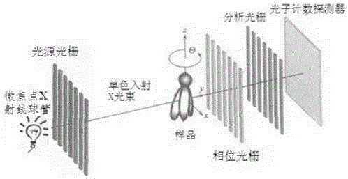
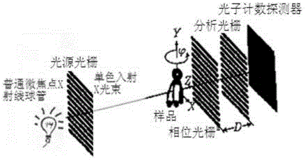
[0061] figure 2 It is a schematic diagram of the overall structure of the X-ray imaging system in one embodiment of the present invention. The X-ray imaging system includes components such as an X-ray source, a light source grating, a sample scanning platform, a phase grating, an analysis grating, a photon counting detector, and a three-dimensional reconstruction system (not shown in the figure). The above light source grating, phase grating and analysis grating have a plana...


PUM


Abstract

The invention discloses an X-ray imaging system and method based on grating phase contrast and photon counting. X-rays become coherent X-rays after being shaped through a light source grating; the coherent X-rays containing phase changes after penetrating through samples form beam-split X-rays through a phase grating; the phase changes of the X-rays are converted into light intensity changes after the X-rays pass an analyzing grating; a photon counting detector is used for recording phase contrast information of the X-rays with different intensities; sectional images based on phase contrast are obtained through a three-dimensional reconstruction system; finally, components and interior fine structure information of the soft tissue samples are obtained. The system and method can be used for detecting soft tissue samples in the pathology department, the radiology department and the scientific research department of a hospital, early lesion information such as small lesions in the tissue samples can be found easily, and the detection rate is greatly increased.

Description

technical field [0001] The invention relates to an X-ray imaging system, in particular to an X-ray imaging system based on grating phase contrast and photon counting technology, and also relates to a method for realizing X-ray imaging by the system, which belongs to the technical field of medical imaging. Background technique [0002] X-rays are particularly penetrating. When X-rays pass through samples made of light elements such as carbon, hydrogen, and oxygen, it is like visible light passing through glass, leaving no observable traces. This is extremely unfavorable to medical diagnosis. For example, for the diagnosis of breast tumors, breast tumors are still lesions composed of light elements in the early stages of development, and absorption contrast imaging is powerless to them. It is not until breast tumors develop into late stages that calcification occurs can be seen in absorption contrast imaging. The best opportunity for treatment has been missed like this, whic...

Claims


Application Information

Patent Timeline
Patent Type & Authority Applications(China)
IPC IPC(8): A61B6/00
Inventor 郑海亮李运祥曹红光
Owner NANOVISION TECHNOLOGY (BEIJING) CO LTD
Why Eureka
Try Eureka