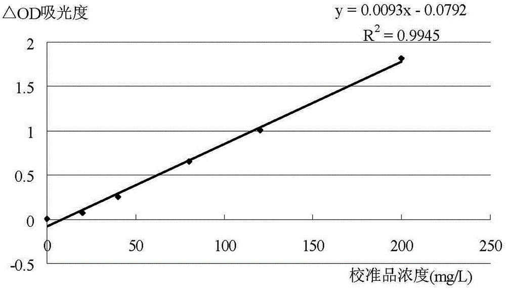
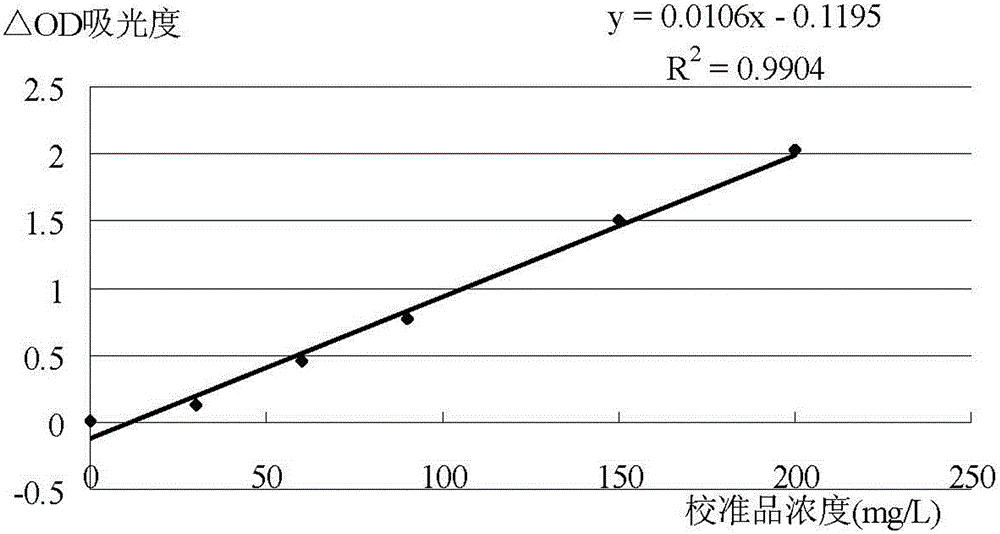
Detection kit and detection method for detecting animal C-reactive protein based on amide group nanometer latex enhanced turbidimetry
A detection kit and nano-latex technology, which is applied in the field of medical immunodiagnosis, can solve problems such as the absence of C-reactive protein, and achieve the effects of good specificity, high sensitivity, and rapid response
- Summary
- Abstract
- Description
- Claims
- Application Information
AI Technical Summary
Problems solved by technology
Method used
Image
Examples
Embodiment approach
[0028] As another preferred embodiment of the present invention, the nanolatex in the C-reactive protein antibody cross-linked amide group nanolatex is covalently bonded to the amino group or carboxyl group of the C-reactive protein antibody through the amide group on the surface. In this preferred case, the combination of the amide group nanolatex and the antibody is through the covalent bonding of the amide group on its surface to the amino group or carboxyl group of the antibody, and there is a bridging chemical arm between the microsphere and the antibody, which reduces the position. The blocking effect not only improves the binding rate of the antibody, but also provides a suitable three-dimensional structure for the antibody, effectively protecting the active area where the antibody binds to the antigen, and improving the sensitivity of the reaction.
[0029] The present invention also provides a method for detecting animal C-reactive protein based on amide group nano-lat...
Embodiment 1
[0040] 1. Preparation of detection kit:
[0041] 1. Take 1ml of polystyrene latex particles (particle diameter: 80nm) with amide groups on the surface and dilute to 100ml with 10mM 2-morpholineethanesulfonic acid buffer, pH=5.0, and the final concentration is 1%.
[0042] 2. Add 0.1% w / v 1-ethyl-(3-dimethylaminopropyl) carbodiimide hydrochloride and 0.1% w / v N-hydroxysuccinamide to activate the latex, and bathe in water at 37°C for 1 hour .
[0043] 3. Centrifuge, remove the supernatant (containing the residual activated cross-linking agent), then add 2 mg of CRP polyclonal antibody for cross-linking reaction, and bathe in water at 37°C for 4 hours.
[0044] 4. Finally, add 1ml of latex blocking solution containing 2% w / v protein BSA, 0.1% w / v sodium azide and 10mmol / L phosphate buffer at pH=7.0 to block the residual activation sites on the latex surface to obtain R2 reagent.
[0045] 5. Take 10ml of 10mmol / L phosphate buffer solution with pH=7.0, add 1%w / v PEG20000, 1%w / v ...
Embodiment 2
[0056] 1. Preparation of detection kit:
[0057] 1. Take 1ml of polystyrene latex particles (particle diameter: 200nm) with amide groups on the surface and dilute to 200ml with 5mM 2-morpholineethanesulfonic acid buffer solution with pH=6.0, the final concentration is 0.5%.
[0058] 2. Add 0.05% w / v 1-ethyl-(3-dimethylaminopropyl) carbodiimide hydrochloride and 0.05% w / v N-hydroxysuccinamide to activate the latex, and bathe in water at 37°C for 1 hour .
[0059] 3. Centrifuge, remove the supernatant (containing the residual activated cross-linking agent), then add 2 mg of CRP polyclonal antibody for cross-linking reaction, and bathe in 37°C water for 3 hours.
[0060] 4. Finally, add 1ml of latex blocking solution containing 1%w / v trehalose, 0.05%w / vProclin-300 and 20mmol / L citric acid-phosphate buffer at pH=8.0 to block the residual activation sites on the latex surface. The R2 reagent is obtained.
[0061] 5. Take 10ml of 20mmol / L phosphate buffer solution with pH=8.0, ad...
PUM
| Property | Measurement | Unit |
|---|---|---|
| diameter | aaaaa | aaaaa |
| concentration | aaaaa | aaaaa |
| concentration | aaaaa | aaaaa |
Abstract
Description
Claims
Application Information
 - R&D
- Intellectual Property
- Life Sciences
- Materials
- Tech Scout
- Unparalleled Data Quality
- Higher Quality Content
- 60% Fewer Hallucinations
Browse by: Latest US Patents, China's latest patents, Technical Efficacy Thesaurus, Application Domain, Technology Topic, Popular Technical Reports.
© 2025 PatSnap. All rights reserved.Legal|Privacy policy|Modern Slavery Act Transparency Statement|Sitemap|About US| Contact US: help@patsnap.com


