
Epithelial-mesenchymal transition model of retinal pigment epithelium and its application

A retinal pigment and epithelial cell technology, applied in the field of retinal pigment epithelium cell epithelial-mesenchymal transition model, can solve the problems that cell models cannot be widely used, and intercellular connections are difficult to be destroyed, and the method is simple and easy to implement. , good repeatability

Active Publication Date: 2018-07-24
TONGJI UNIV
View PDF1 Cites 0 Cited by
  • Summary
  • Abstract
  • Description
  • Claims
  • Application Information

AI Technical Summary

Problems solved by technology

In this model, RPE cells were treated with 2mMEGTA for 48 hours, and after the intercellular connection was interrupted, it was found that the cells at the edge had motility and migration ability, and the morphology also developed towards mesenchymal cells, but the disadvantage was that the intercellular connection in the center was difficult. is destroyed, so no EMT transition occurs
Therefore, this cell model cannot be widely used

Method used


Image

  • Epithelial-mesenchymal transition model of retinal pigment epithelium and its application
  • Epithelial-mesenchymal transition model of retinal pigment epithelium and its application
  • Epithelial-mesenchymal transition model of retinal pigment epithelium and its application

Examples


Embodiment 1

[0037] Cell culture:

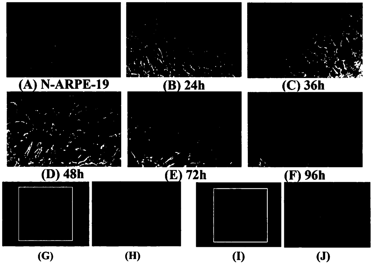
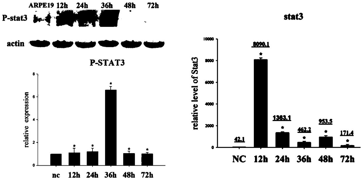
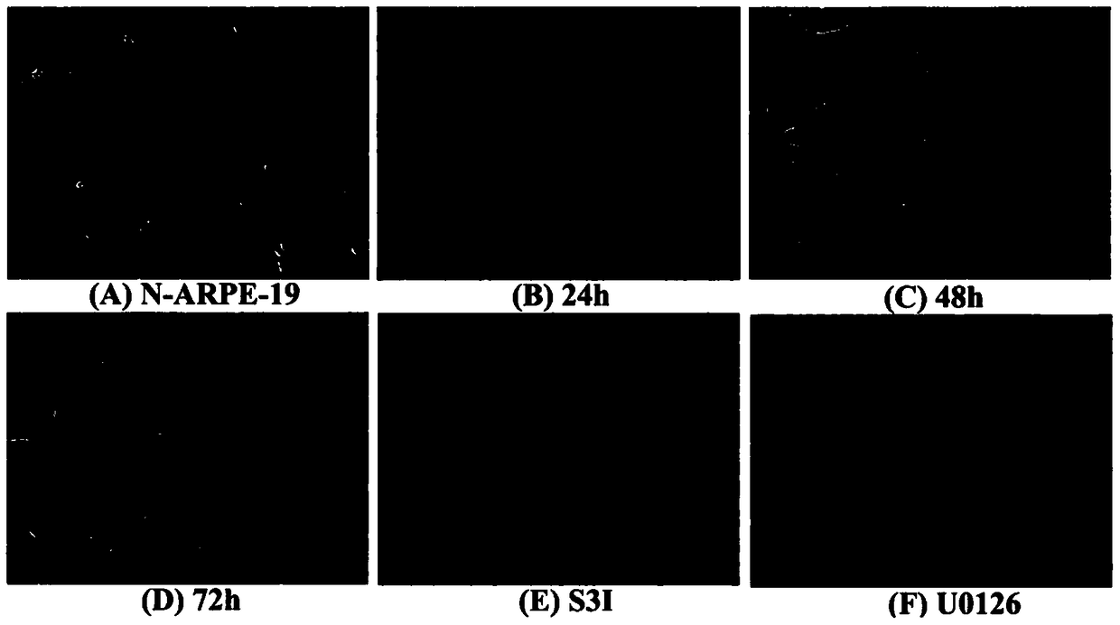
[0038] The culture of ARPE-19 cells was divided into two parts: in the cell dish and in the EMT model. When cultured in a cell dish, culture in DMEM-F12 medium containing 10% fetal bovine serum (FBS) and 1% penicillin-streptavidin double antibody. Cells per dish (10cm culture dish, cell density is 1×10⁷cells / dish). When doing EMT model, ARPE-19 cells were digested with trypsin, and then cultured in serum-free DMEM-F12 medium containing N2 and non-essential amino acids (NEAA) in a bacterial dish, at 12h and 24h respectively After 36h, 48h, and 72h, cell samples were collected or dissolved in trizol, and detected by Q-PCR, ELISA, Western blot and other methods. The cells in two dishes were added with S3I (Stat3 inhibitor) and U0126 respectively at 24h, and the samples were collected at 48h. In the experiment of detecting stat3 activation, the pSTAT3-TA-luc reporter gene plasmid was first transfected into ARPE-19 with lipofectaminer 2000, and after 48 ho...

Embodiment 2

[0053] To investigate the inhibitory effect of mir-24 on the EMT of ARPE-19.

[0054] 1. Cell culture

[0055] The main body of cell culture is mir-24-ARPE-19 cells, which are divided into two parts, one of which is to verify the effect of mir-24 on the EMT, proliferation and migration of ARPE-19 cells, as well as the effect of stat3 signaling pathway. One part is to clarify the targeting effect of mir-24 and inflammatory factor CHI3L1.

[0056] The details are as follows: Validation of the effect of mir-24 on the EMT of ARPE-19 cells: cultured in serum-free DMEM / F12 containing N2 and NEAA, petri dish. To verify the effect of mir-24 on the proliferation and migration of ARPE-19 cells: culture in DMED / F12 culture medium with 10% fetal bovine serum, cell dish, scratch after 24 hours, replace with serum-free culture medium and continue to culture. To verify the effect of mir24 on the stat3 signaling pathway in the EMT process of ARPE-19 cells: cultured in DMED / F12 medium with 1...


PUM


Abstract

The invention relates to a retinal pigment epithelial cell epithelial-mesenchymal transition model and its application. ARPE-19 cells were digested with trypsin, then transferred to serum-free DMEM-F12 medium containing N2 and non-essential amino acids, and cultured in Petri dishes to obtain retinal pigment epithelial cells epithelial-mesenchymal The mesenchymal transition model; this model is used to study the molecular mechanism of epithelial-mesenchymal transition in retinal pigment epithelial cells and to find the intervention target of epithelial-mesenchymal transition. Compared with the prior art, the method of the invention is simple and easy to implement and has good repeatability.

Description

technical field [0001] The invention relates to a cell model, in particular to a retinal pigment epithelial cell epithelial-mesenchymal transition model and its application. Background technique [0002] Proliferative vitreoretinopathy (PVR) is the most important reason for the failure of rhegmatogenous retinal detachment surgery. Because of its refractory and recurrence, it has always been a research hotspot by scholars from all over the world. The incidence of PVR is 5%- 20%. PVR is a proliferative disease. The basic pathological process of PVR is that after a retinal hole occurs, the blood-retinal barrier is destroyed, and the proteins and active mediators (such as transforming growth factor (TGF)-β, and fibroblast growth factor (FGF)) in the vitreous are released, and the retinal pigment epithelial cells (retinal pigment epithelium) Epithelial (RPE) exposure, loss of contact with photoreceptors, RPE dissociation, migration, cytokines stimulate RPE proliferation, cell p...

Claims


Application Information

Patent Timeline
Patent Type & Authority Patents(China)
IPC IPC(8): C12N5/071C12N5/10
Inventor 吕立夏徐国彤
Owner TONGJI UNIV
Learn More