A method for double staining of bovine in vitro fertilized blastocysts

A double staining and in vitro fertilization technology, which is applied to the quality evaluation of bovine in vitro fertilized blastocysts. It is simple and fast in the field of double staining of bovine blastocysts. It can solve the problems of long dyeing time, time-consuming, and affecting the dyeing effect. Effect of shortening dyeing time, reducing required time, and reducing dyeing time

Inactive Publication Date: 2017-10-03
天津市农业科学院
View PDF4 Cites 0 Cited by
  • Summary
  • Abstract
  • Description
  • Claims
  • Application Information

AI Technical Summary

Problems solved by technology

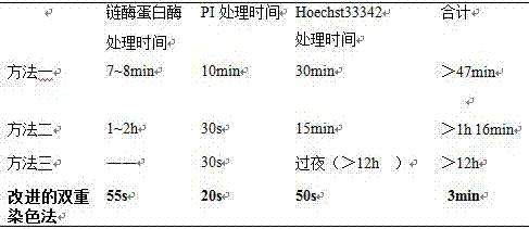
The current blastocyst double staining method is established on the basis of the double staining of blastocysts by Thous et al. using TritonX-100, but the biggest problem is that the procedure is cumbersome, and each step of staining takes a lot of time. The whole staining procedure takes at least 47 minutes. Above, so the staining time is long (Li Ruiqi, Sang Runzi. The effect of fructose on the early development of bovine embryos was tested by double staining of blastocysts[J]. China Journal of Animal Husbandry, 2010 (9): 23-25; Li Rong, Liu Ying, Zhao Xingbo, et al. Application of serum-free medium IVD101 and G1 / G2 in bovine somatic cell nuclear transfer[J]. Journal of Animal Husbandry and Veterinary Medicine, 2008,39(11):1487-1492; Thouas GA, Korfiatis N A, French A J, et al. Simplified technique for differentialstaining of inner cell mass and trophectoderm cells of mouse and bovineblastocysts[J]. Reproductive biomedicine online, 2001, 3(1): 25-29.)
[0004] Usually when evaluating the quality of blastocysts, due to the different ages and developmental stages of embryos, it is necessary to classify the embryos and dye them in multiple batches, so the dyeing takes a long time, the efficiency is low, and the effect of dyeing may be affected

Method used


Image

  • A method for double staining of bovine in vitro fertilized blastocysts
  • A method for double staining of bovine in vitro fertilized blastocysts
  • A method for double staining of bovine in vitro fertilized blastocysts

Examples


Embodiment 1

[0027] 1. Materials and Methods

[0028] 1.1 Collection of oocytes

[0029] Cattle ovaries were taken from the slaughterhouse, placed in 37°C normal saline with penicillin and streptomycin, transported back to the laboratory for 2-3 hours, removed the surrounding adipose tissue, and then treated with normal saline with penicillin and streptomycin Rinse 5-6 times, use the egg-collecting fluid suction method to absorb follicles with a diameter of 2-8 mm, and select naked eggs, semi-naked eggs, and cumulus-oocyte complexes with three or more layers of cumulus according to different needs of the experiment (Cumulus oocyte complexes, COCs) were used for experiments;

[0030] Egg collection fluid: TCM-199 (tissue culture medium) + 5% FBS (fetal bovine serum) + 30µg / ml Heparin (sodium heparin) + 4.766g / l Hepes (4-hydroxyethylpiperazineethanesulfonic acid)

[0031] 1.2 In vitro maturation culture

[0032] Wash the collected oocytes with maturation solution for 3 times, and then tr...

Embodiment 2

[0048] 1. Materials and Methods

[0049] 1.1 Collection of oocytes

[0050] Cattle ovaries were taken from the slaughterhouse, placed in 37°C normal saline with penicillin and streptomycin, transported back to the laboratory for 2-3 hours, removed the surrounding adipose tissue, and then treated with normal saline with penicillin and streptomycin Rinse 5-6 times, use the egg-collecting fluid suction method to absorb follicles with a diameter of 2-8 mm, and select naked eggs, semi-naked eggs, and cumulus-oocyte complexes with three or more layers of cumulus according to different needs of the experiment (Cumulus oocyte complexes, COCs) were used for experiments;

[0051] Egg collection fluid: TCM-199 (tissue culture medium) + 5% FBS (fetal bovine serum) + 30µg / ml Heparin (sodium heparin) + 4.766g / l Hepes (4-hydroxyethylpiperazineethanesulfonic acid)

[0052] 1.2 In vitro maturation culture

[0053] Wash the collected oocytes with maturation solution for 3 times, and then tr...


PUM


Abstract

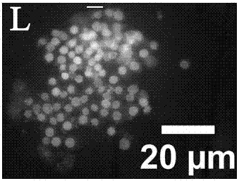
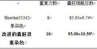
The invention discloses a double-staining method for bovine in vitro fertilized blastocysts, which comprises treating the bovine in vitro fertilized blastocysts cultivated to 7-9 days with 0.5% pronase for 55 seconds, and then washing them in PBS for 2 to 3 times. Stain in 100µg / ml PI (1%TritonX‑100 / PBS) for 20s, wash in PBS for 2-3 times, transfer to 45µg / ml Hoechst33342 for staining for 50s, wash in PBS for 2-3 times, and press. After pressing, the tablets were observed and photographed under an inverted fluorescent microscope under ultraviolet light. The nuclei of the blastocyst inner cell mass (ICM) are blue, and the nuclei of trophoblast (TE) cells are red. The staining method can be completed in 3 minutes, while the previous report required at least 47 minutes. Therefore, the time required for double staining of blastocysts is greatly reduced, and the efficiency is improved, so that the quality of bovine blastocysts can be quickly evaluated.

Description

technical field [0001] The invention belongs to the biological technical field of bovine in vitro fertilization, and relates to a method for evaluating the quality of bovine in vitro fertilized blastocysts, in particular to a simple and rapid double-staining method for bovine blastocysts. Background technique [0002] Since the successful establishment of the cattle IVP system, people have tried to improve the system from different angles, [0003] For example, changing the culture system, adding lipid metabolic regulators, adding cAMP regulators (Cyclic adenosine monophosphate (cAMP) modulators), etc., but the quality of bovine in vitro embryos is still poor compared with in vivo embryos, not only the cyst The number of blast cells is less and the pregnancy rate of transfer is not high. This makes people realize the necessity of improving the quality of bovine embryos, so the inspection of embryo quality has gradually become one of the important indicators to evaluate the ...

Claims


Application Information

Patent Timeline
Patent Type & Authority Patents(China)
IPC IPC(8): G01N1/30
Inventor 刘海军林峰黄承俊
Owner 天津市农业科学院
Why Eureka
Try Eureka